Radioactivity in Medicine
Brook Edgar & Hannah Shuter
Teachers


Contents
Explainer Video
Radiotherapy
External Radiotherapy
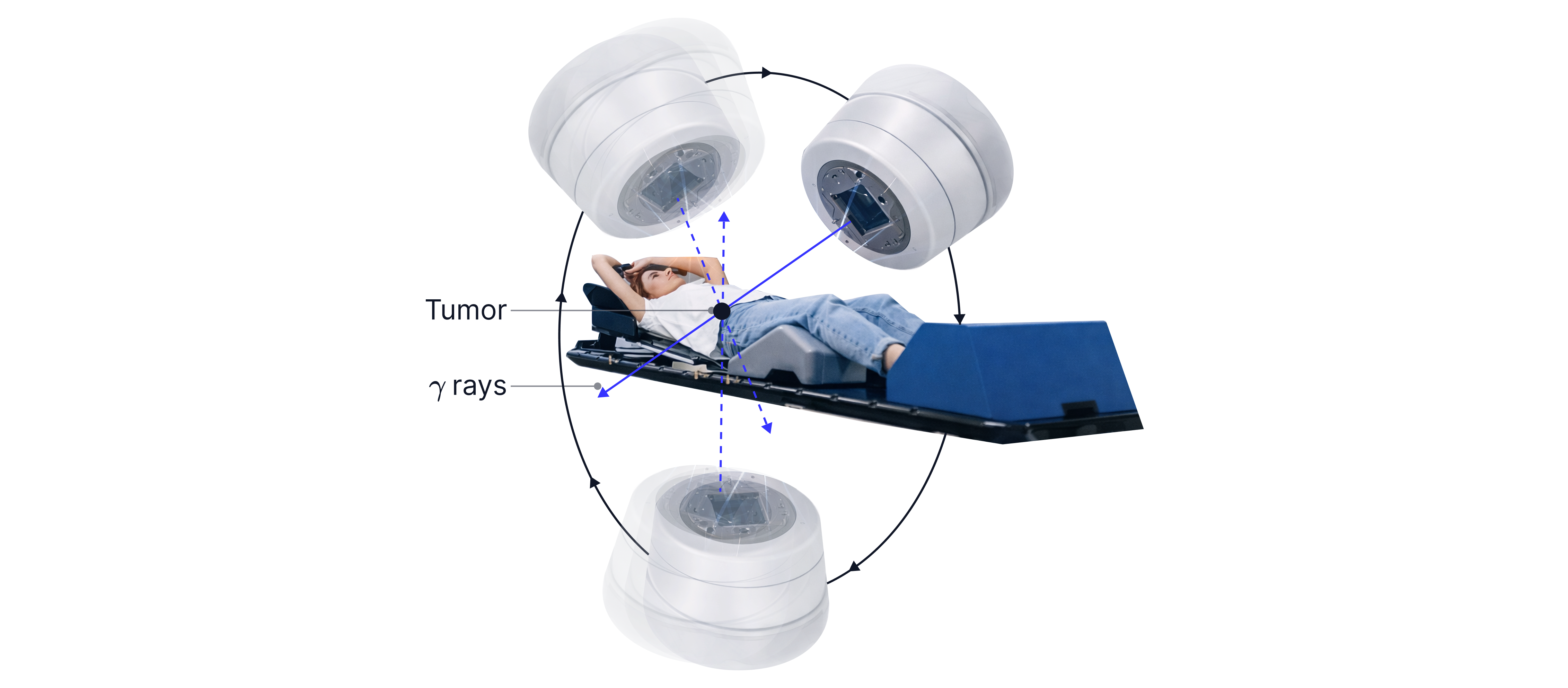
Radiotherapy uses focused beams of high-energy ionising radiation (gamma rays or X-rays) to damage the DNA of cancer cells, preventing them from dividing and causing them to die.
To prevent damage to healthy cells, imaging with CT or MRI is used to identify the exact position of the tumour, so the radiation can be targeted precisely with narrow, collimated beams. The source rotates around the patient so that many beams enter from different directions. Each beam delivers only a small dose to the healthy tissue it passes through, but the beams all intersect at the tumour, where the total dose is highest. Lead shielding is also used to protect nearby healthy tissues and organs.
Internal radiotherapy
Brachytherapy is a form of radiotherapy in which sealed radioactive sources are placed inside or very close to a tumour.
Radioactive implants may use beta-emitting isotopes because beta particles have a short range and are highly ionising. This delivers a high dose to nearby cancer cells while limiting damage to healthy tissue further from the implant.
Advantages:
Healthy tissue further away is less affected than by external radiotherapy
Can deliver higher doses locally
More effective than external radiotherapy for some tumours
Worked Example:
Internal radiotherapy can be used to treat cancers that are confined to a small area of the body. A sealed radioactive source is implanted close to the tumour.
Explain one advantage of using a beta-emitting source instead of a gamma-emitting source for this treatment.
Answer:
Beta radiation is more ionising than gamma radiation, so will be more effective at treating the tumour.
OR
Beta is less penetrating than gamma, so it will cause less damage to surrounding healthy tissue.
Radionuclide Imaging
Radionuclide imaging – imaging the body using radioactive tracers that accumulate in specific organs or tissues.
A radionuclide is an unstable atomic nucleus that undergoes radioactive decay, emitting ionising radiation.
Radionuclide imaging uses (mainly) gamma-emitting radioactive tracers to investigate specific regions of the body. A radioisotope is bound to a substance that follows a particular biological pathway - e.g. attached to red blood cells to image blood flow. As the radioisotope decays, it emits (mainly) gamma rays that escape the body (highly penetrating) and are detected by a gamma camera outside the body. A computer processes the detected gamma rays to produce images showing the distribution of the tracer within the body. 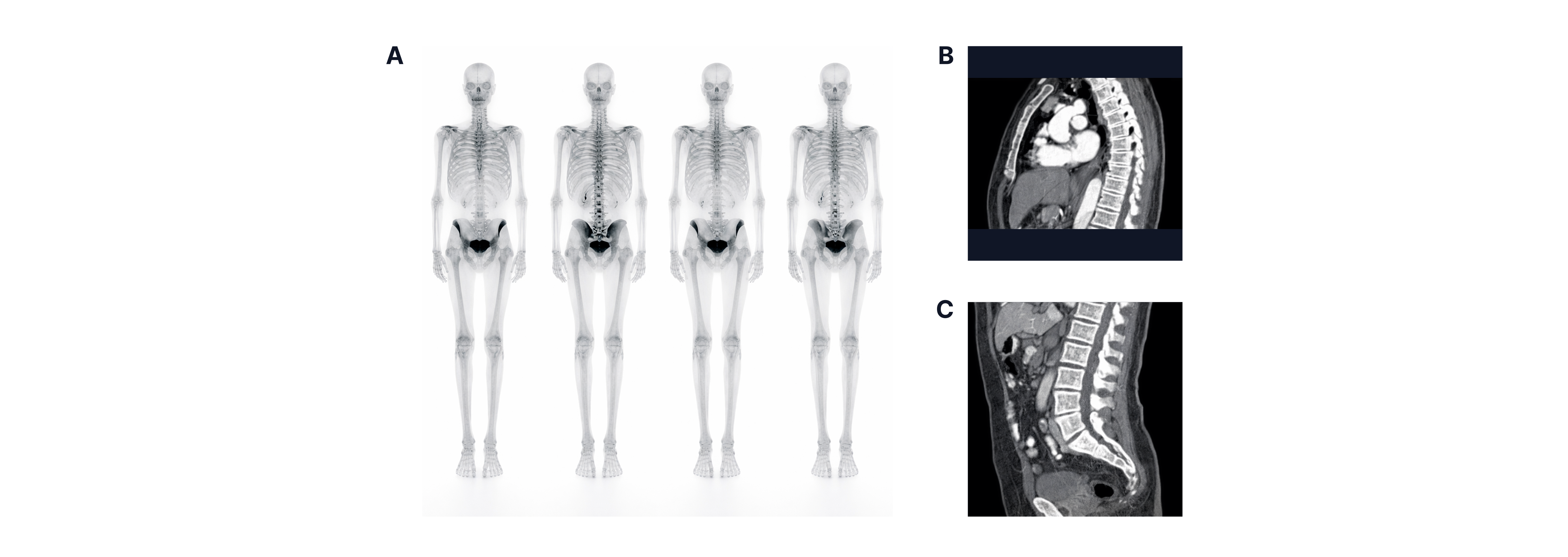
Alpha and beta radiation have lower penetration and are more ionising than gamma radiation; they are not used because they would be absorbed within the body, increasing the radiation dose to healthy tissues.
Common Medical Radioisotopes
Isotope | Technetium-99 | Iodine-131 | Indium-111 |
Emits | Pure gamma | Beta and gamma | Pure gamma |
Energy | |||
Physical Half-life | |||
Uses | Bone scans, blood flow imaging, heart and kidney imaging. | Thyroid imaging and cancer treatment | Labelled white blood cells and detecting infections |
Other information | Easily prepared on site, short half-life reduces patient dose | Iodine is naturally absorbed by the thyroid | Quite expensive |
Production of Technetium-99m
Technetium- has a short half-life (6 hours), so it would decay significantly during transport. Instead, it is transported as molybdenum-99 which decays into technetium-as molybdenum-99 has a much longer half-life of about 66 hours (approximately 2.75 days).
Molybdenum-99 is strongly bound to an aluminium oxide inside the molybdenum-technetium generator, whereas the technetium- produced is not. Saline can be passed through to wash the technetium- out, allowing hospitals to extract technetium- at regular intervals/when needed.
Effective Half-Life
When dealing with radioactive tracers, we need to take into account not only the physical half-life of the isotope but also how quickly it is removed from the body (its biological half-left to determine how much radiation the patient receives.
Definitions:
Physical half-life = the time taken for the activity (number of nuclie/mass) of a radioactive source to halve due to radioactive decay only. Determines how long the tracer remains radioactive enough to produce a useful image.
Biological half-life = the time taken for half of the radioactive substance to be removed from the body by biological processes (e.g. excretion or metabolism), ignoring radioactive decay. Determines how long the tracer stays in the organ being imaged.
Effective half-life = the time taken for the activity of the radionuclide in the body to halve due to both radioactive decay and biological removal. This determines the actual radiation dose received by the patient.
Formula:
= effective half-life
= physical half-life
= biological half-life
Worked Example:
Technetium- is commonly used as a radioactive tracer in medical imaging. After being injected into a patient, its activity decreases due to both radioactive decay and the body's natural elimination processes.
The physical half-life of technetium is .
The biological half-life of technetium in a patient's body is .
Calculate the effective half-life of technetium in the patient's body.
Answer:
Worked Example:
Technetium- must be produced close to the hospital where it is used, whereas iodine-131 can be transported over much longer distances.
Explain this difference in terms of the half-lives of the two isotopes.
Explain why technetium- is generally considered to be safer for patients than iodine-131 when used as a radioactive tracer.
Answer:
Technetium- has a half-life of around , whereas iodine-131 has a half-life of around .
This means technetium would lose a significant proportion of its activity during transportation, so must be produced on site.
Technetium has a shorter half-life than iodine, so its activity declines rapidly after a scan.
This means the patient receives a lower dose of radiation from technetium than from iodine.
Remember: When calculating half-life, you may also be asked to determine the decay constant and the activity of a source. Make sure to revisit the 'Radioactive Decay' page in Topic 11 to review how to use these equations.
PET Scanners
PET (Positron Emission Tomography) is an imaging technique in which a patient is injected with a positron-emitting radionuclide tracer (a beta-plus emitter). These emitted positrons almost immediately collide with electrons in the surrounding tissue. They annihilate each other, converting their mass into the energy of two gamma photons, which are emitted in opposite directions. The gamma photons emitted are detected by a ring of gamma cameras (see below) around the patient, and the differences in detection times allow a computer to pinpoint exactly where the photons originated. By detecting many such events, the computer reconstructs a detailed image of the tracer's distribution within the body.

Image formation depends on metabolic activity. Metabolically active cells are those carrying out chemical reactions at a high rate, so they require more energy and nutrients. Cancer cells often have a high metabolic rate. More metabolically active cells take up more radiotracer (it behaves like glucose), resulting in more positron emissions and, therefore, more annihilation events. This produces more gamma photons, so these regions appear brighter on the PET image.
PET Advantages
Measures metabolic activity, not just anatomy
Can detect tumours early, as cancer cells have a high metabolic rate
Shows the extent (spread) of cancer/tumour
Can measure brain activity and heart function, as tissues in the brain and heart have a high metabolic rate
PET Disadvantages
Uses ionising radiation
Long scan time, as the radiotracer must be distributed throughout the body before imaging
Large and expensive equipment
lower resolution than CT or MRI, so PET scans are often combined with CT scans to provide detailed anatomical images and more accurate tumour localisation.
Gamma Camera
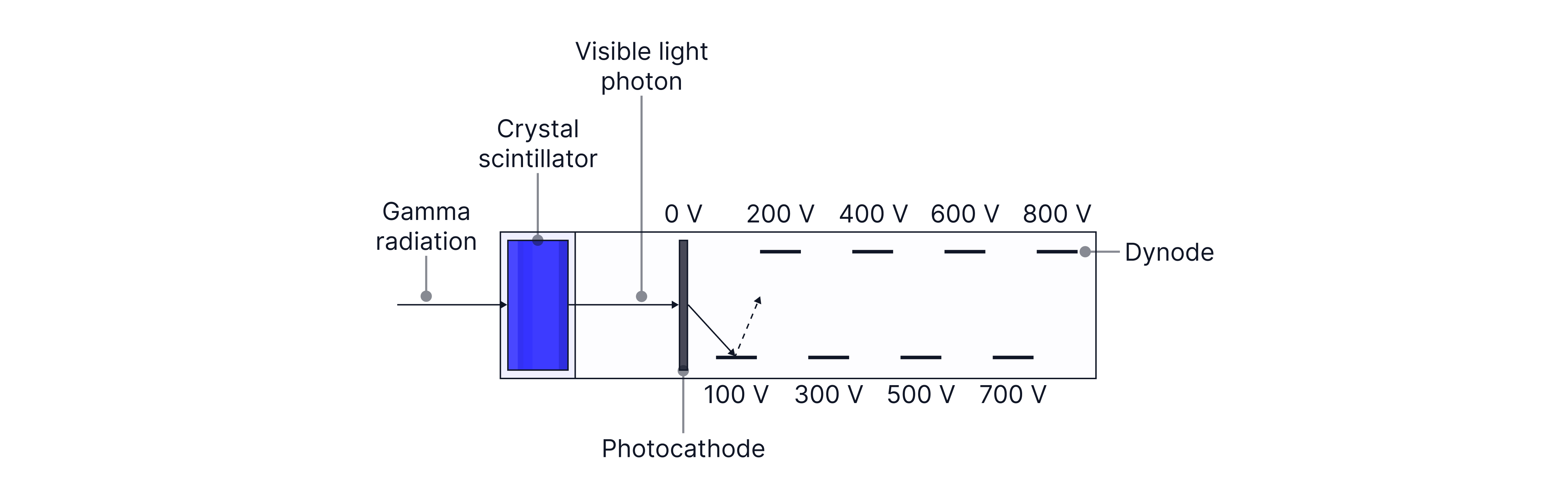
A gamma camera uses a lead collimator, a scintillator crystal and a photomultiplier tube (PMT) to detect gamma rays - as shown in the diagram below. A gamma ray emitted by a patient first enters the lead collimator. The purpose of the collimator is to absorb off-axis gamma rays to improve image resolution.

Only near-parallel gamma photons reach the scintillator crystal, which absorbs energy from the gamma ray and emits visible light. This light then hits a photocathode, which releases electrons by the photoelectric effect. These electrons are accelerated towards a series of dynodes (shown below). Each electron striking a dynode emits four secondary electrons, multiplying the number of electrons at each stage. This amplification produces a measurable electrical pulse, which is processed by a computer to determine the location and intensity of the detected gamma ray.
Worked Example:
A patient is injected with a radioactive tracer before undergoing a scan with a gamma camera.
When a gamma photon strikes the scintillation crystal, a flash of visible light is produced.
Explain how the photomultiplier tube converts this light into a much larger electrical signal.
Answer:
Light strikes the photocathode and it emits an electron
The electron is accelerated towards the dynodes
Each electron collision with a dynode produces 4 more electrons
Worked Example:
PET scanners use the difference in detection time to pinpoint the position of the radioisotope. Determine the difference in distance to the detector given by a time difference of .
Answer:
Remember that PET scanners emit gamma rays, which travel at . Therefore:
Practice Questions
A radioactive tracer used in medical imaging undergoes a decay that produces a positron.
As a result, two detectors positions on opposite sides of the patient record gamma photos at the same time.
Describe the sequence of events that leads to the production of these gamma photons.
-> Check out Hannah's video explanation for more help.
Answer:
Positron meets and electron and annihilates
Mass of the positron and the electron is converted into the energy of the gamma photons
Gamma photons must move in opposite directions to conserve momentum
A radioactive tracer is used in medical imaging and detected using a gamma camera.
The tracer used is indium-111 which has a physical half-life of .
A patient is injected with indium-111 with an initial activity of .
For this patient, the biological half-life is .
For radiation protection reasons, the patient must remain in hospital until the activity in their body falls below .
Determine whether the patient can be safely discharged after .
-> Check out Hannah's video explanation for more help.
Answer:
First, calculate the effective half-life:
Now use this to find the decay constant of indium-111:
Now we can calculate the activity after 4 days:
Therefore, the patient is safe to be discharged after .