X-Ray Imaging
Brook Edgar & Hannah Shuter
Teachers


Contents
Explainer Video
Production of X-Rays
X-rays are high-energy ionising EM waves. They are used to image the body because different tissues absorb X-rays by different amounts. In traditional radiography, X-rays passing through the body darken a photographic film, producing the familiar X-ray image of structures such as broken bones. X-rays transmitted through the body can also form images by being converted into visible light or electrical signals, which are then processed by a computer. X-rays are also used in CT scans to produce detailed cross-sectional images of the body.
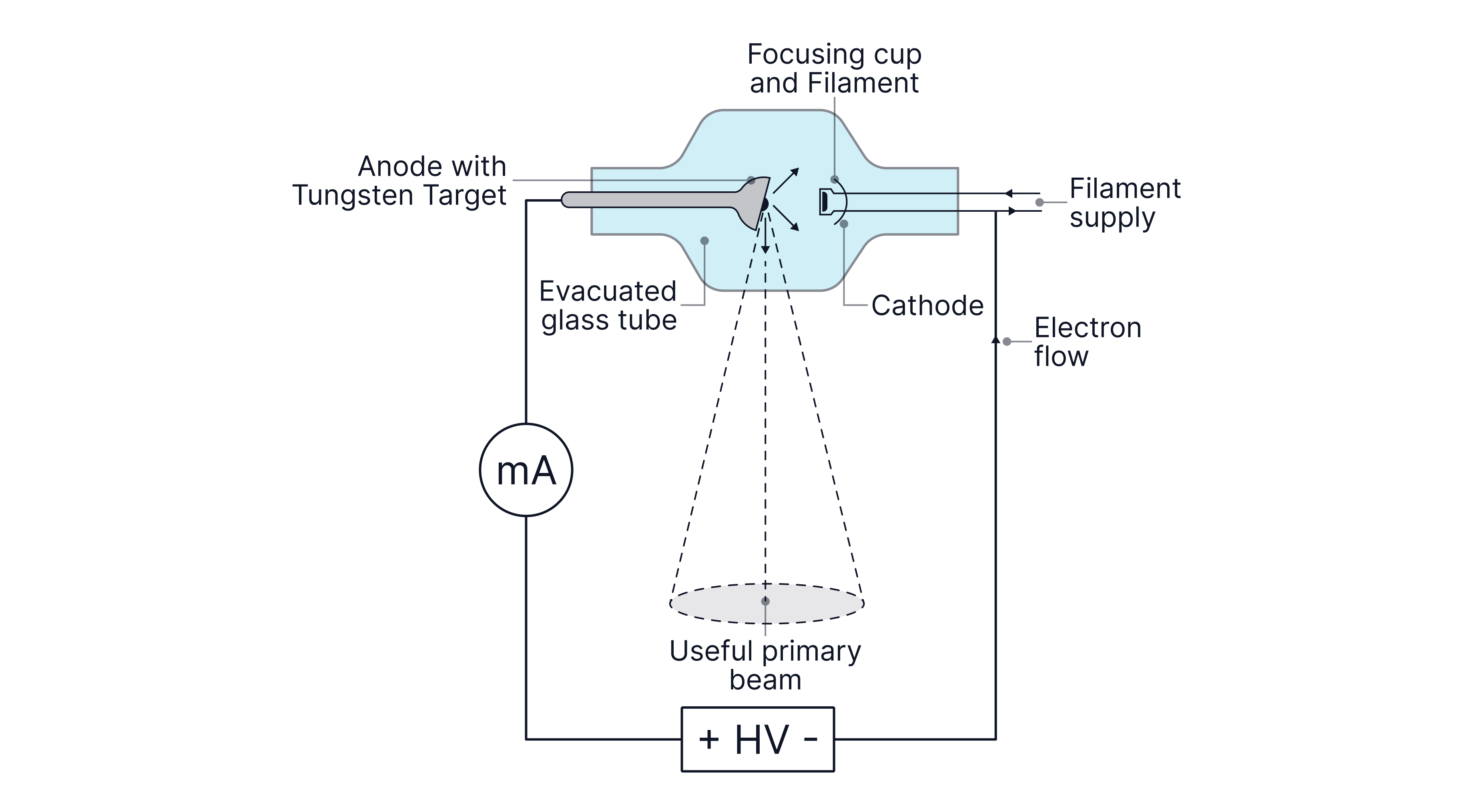
X-rays are produced in an X-ray tube:
-> A heated filament (cathode) releases electrons by thermionic emission
-> Electrons are accelerated through a large potential difference ( )
-> Electrons collide with a metal target (anode), usually tungsten, producing X-ray photons. Tungsten is used because of its high melting point and atomic number. Only of the electrons' kinetic energy is converted into photons, meaning is converted into heat. The anode is large and rotated, so the heat is spread over a large surface area, reducing the temperature rise, allowing X-rays to be geberatedn for longer. It is also bevelled (angled) to produce a larger target area but a small focal spot (source area) to produce a sharp image (better resolution).
X-rays can be produced by the anode by either:
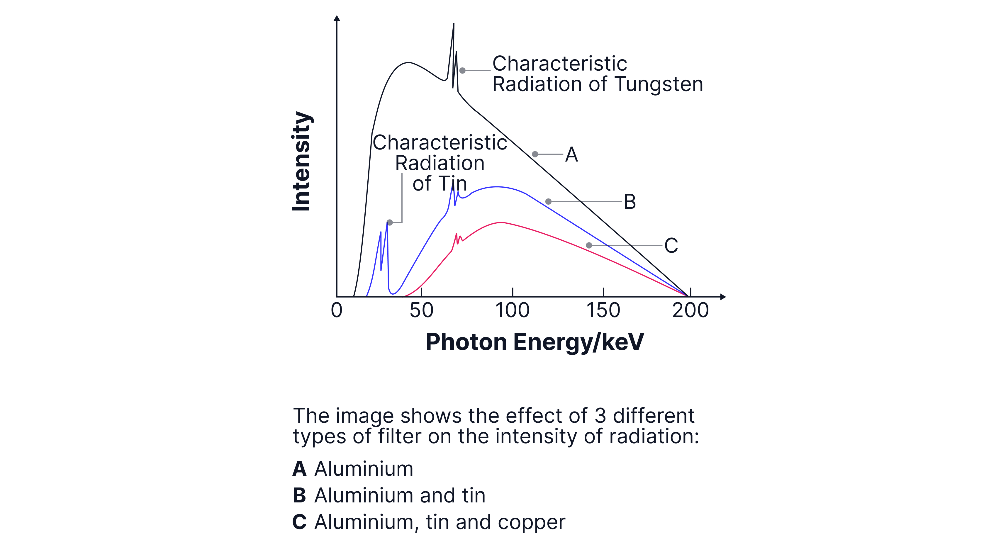
Bremsstrahlung radiation -> When a charged particle is accelerated or decelerated, it emits EM radiation. Fast-moving electrons are attracted by the tungsten nucleus's large positive charge, causing them to change direction and decelerate. The kinetic energy lost by the electrons is emitted as X-rays. Because electrons can lose different amounts of energy in each interaction, a continuous spectrum of X-ray energies is produced.
Characteristic X-rays -> Incident electrons have enough energy to eject inner-shell electrons from tungsten atoms, causing outer-shell electrons to fall down into the vacancies, emitting X-rays. Since the energy levels in the atom are discrete, the emitted X-rays have discrete (specific) energies, producing characteristic X-ray lines.

Note:
-> If the accelerating voltage is increased, it increases the maximum photon energy and the intensity of the X-ray radiation, as more energetic electrons are more efficient at producing X-rays.
-> If the tube current is increased, more electrons per second travel from the cathode to the anode, but the energy of each electron remains unchanged; therefore, only the intensity of the X-rays increases as more photons are emitted each second.
Radiation Dose and Safety
X-rays are ionising radiation. Ionisation can damage DNA, which can cause cell mutations, damage healthy tissues and increase cancer risk. The biological effect of radiation dose is measured in sieverts (Sv). To reduce the risks associated with ionising radiation, several dose reduction methods are used when taking X-ray images:
Minimising exposure times - reduces the total radiation dose.
Lead shielding - patient wears a lead apron to protect tissues not being imaged
X-ray tube cased in lead - produces X-rays in a directed beam, resulting in less exposure to surrounding tissues
Reduce the distance from the source to the patient - reduces divergence in the beam
Keeping the patient still - prevents the image from getting blurred and thus ensures image repeats are not needed
Using a lead grid between the patient and the film - the lead absorbs scattered X-rays, preventing the image from getting blurred and repeated images from being needed.
Filtering low-energy X-rays using aluminium - low-energy X-rays are more likely to be absorbed by a patient's skin, increasing the radiation dose without improving image quality. Aluminium, copper or tin filters are used to remove these.
Worked Example:
In an X-ray tube, high-energy electrons strike a tungsten target. Explain how these interactions lead to the formation of both a continuous X-ray spectrum and characteristic X-ray lines.
Answer:
The continuous spectrum is due to the decelerating electrons transferring their kinetic energy to x-ray photons
Electrons are decelerated by different amounts so a continuous spectra is emitted.
The characteristic spectra is due to the accelerated electrons knocking inner electrons from atoms.
Outer electrons drop down to fill the gaps left behind and emit x-ray photons of specific energies.
Worked Example:
Discuss whether an X-ray image of a chest or an X-ray image of a leg is likely to be sharper when exposed to the same X-ray source.
Answer:
The x-ray image of the leg would be sharper as it is thinner than the chest so will be closer to the x-ray plate.
Can keep a leg still but it is difficult to keep the chest still as the heart will be beating.
The chest isn't uniformly thick so the front and back will be unable to be focussed at the same time
Attenuation of X-Rays
Attenuation is the reduction in X-ray intensity as it passes through matter. It depends on the material's thickness, density, and atomic number.
Formula:
The higher the density of the material → larger → greater attenuation → the greater the percentage of the X-rays will be absorbed.
Attenuation is important, as different tissues will absorb X-rays by different amounts, providing contrast in the images, allowing different tissues to be identified:
Bone has a high density and a high atomic number, so it absorbs (or attenuates) a large proportion of the X-rays. They appear white on X-ray images.
Air absorbs a very small proportion of the X-rays. It appears black on the X-ray image.
Soft tissues, such as muscle and organs, absorb a smaller proportion of X-rays. They appear grey on the X-ray image.
However, sometimes tissues have similar absorptions, so a contrast medium is used. Contrast media contain elements with high atomic numbers to increase attenuation in specific organs, thereby improving contrast between tissues. A common example is a barium meal, which is used to image the digestive system.
Half-Value Thickness
The half-value thickness is the thickness of a material needed to reduce intensity by .
Formula:
The formula above is not given on the data sheet but can be derivied easily as, .
Mass Attenuation Coefficient
If we want to compare different materials, we sometimes use the mass attenuation coefficient, which also takes the density of the material into account.
Formula:
Worked Example:
X-rays are used in airport security scanners to inspect luggage. As the X-ray beam passes through a metal plate, its intensity decreases because some of the X-rays are absorbed.
A beam of X-rays passes through of aluminium. The linear attenuation coefficient of aluminium is .
Calculate the fraction of the original X-ray intensity that passes through the aluminium.
Answer:
Note - there is no need to change the units of thickness and linear attenuation coefficient if they are in the same units as and will cancel out.
Worked Example:
X-rays are used in hospitals to produce images of bones. A sheet of aluminium is used as part of the shielding around an X-ray machine.
The linear attenuation coefficient of aluminium for the X-rays is .
The density of aluminium is .
Calculate the half-value thickness of the aluminium.
Calculate the mass attenuation coefficient of the aluminium.
Answer:
Image Detection Methods
Traditional X-ray images were produced by photographic film darkening when exposed to X-rays. Image quality improves with increased exposure time to enhance contrast and with increased X-ray energy to improve penetration. However, both methods increase the radiation dose to the patient, so other techniques have been developed to improve image quality without increasing exposure time.
1. Intensifying Screen
X-rays pass through the patient and strike intensifying screens containing crystals that fluoresce when exposed to X-ray radiation. The electrons in the crystals absorb energy from the X-rays, become excited and then de-excite, emitting visible photons.
A double-sided photographic film is placed between intensifying screens, which are much more sensitive to visible light than X-ray photons
The purpose of the two screens is to reduce X-ray dose/exposure time. Using two screens ensures that as much of the transmitted X-ray energy as possible is converted into visible light. The first screen converts X-rays into visible photons, while the back screen converts X-rays that pass through the film into light on the other side, increasing the film's exposure, allowing a shorter exposure time/lower X-ray dose while still producing a sufficiently bright image.

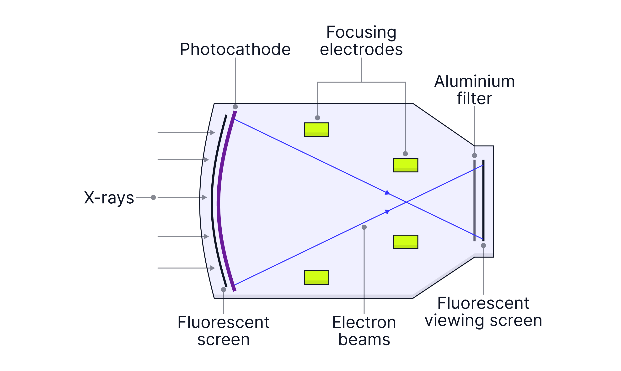
2. Image Intensifier (fluoroscopy) -> Used for real-time/moving images (observing blood flow)
X-rays pass through the patient and strike a fluorescent screen, producing visible light.
Light hits a photocathode, which emits electrons (photoelectric effect)
Electrons are accelerated and focused by electrodes
They strike an output fluorescent screen, producing a much brighter visible image, by up to . Because the image is so much brighter, the X-ray tube doesn't need to produce as many X-rays; normally, if the image were too dim, you would increase the X-ray intensity. A brighter image means less exposure time, thereby reducing the patient's radiation dose.
The image is viewed using a camera and displayed on a monitor
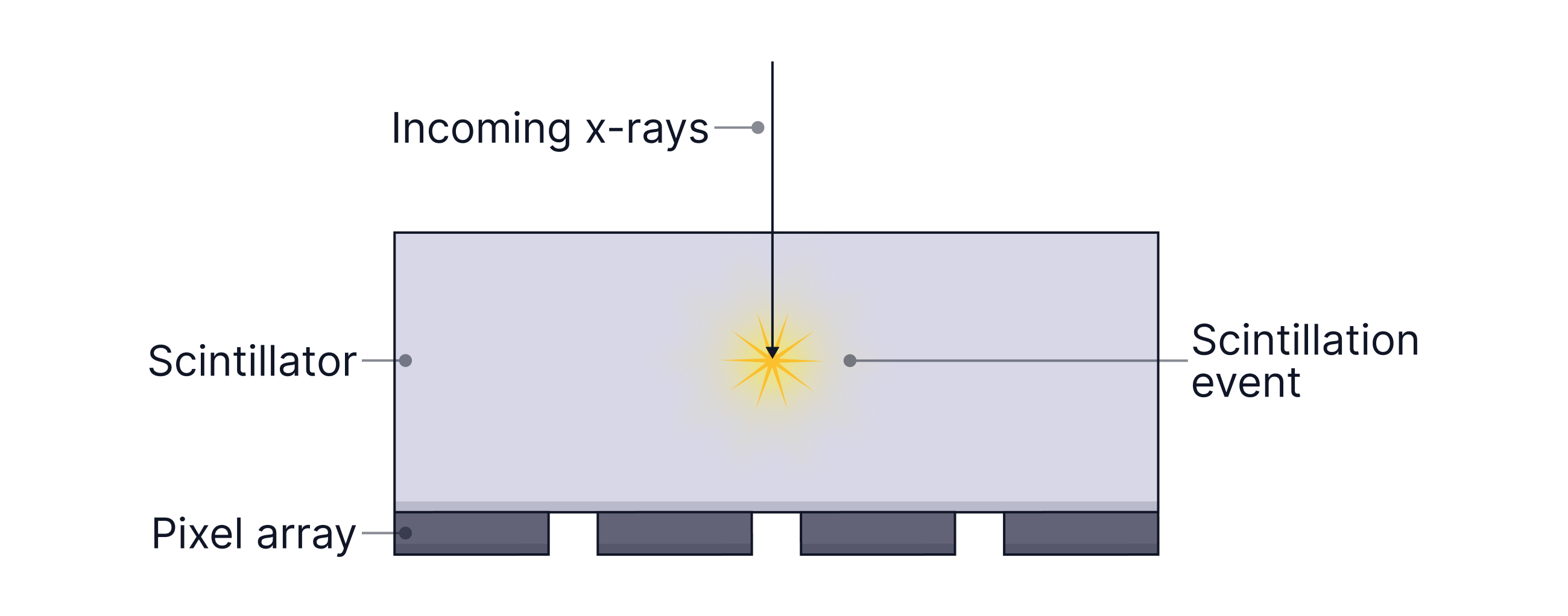
3. Flat Panel (FTP) Digital Detectors -> Very sensitive. Requires less radiation to generate an image
X-rays pass through the patient, hitting a flat panel detector containing a scintillator
A scintillator absorbs ionising radiation -> X-rays, and emits visible light (electrons in the scintillator are excited and then de-excite, emitting visible photons). A photodiode then converts the visible photons into electrical signals. Each photodiode stores charge → pixel.
The signals can be amplified and processed by a computer to produce a digital X-ray. The computer can adjust brightness and contrast after the image is taken, allowing a good-quality image to be obtained with a lower X-ray dose.
Worked Example:
A patient is undergoing a barium swallow examination to investigate a suspected narrowing of the oesophagus. During the procedure, the radiographer needs to observe the movement of the barium solution as the patient swallows.
Explain whether an image intensifier or an intensifying screen should be used to produce the X-ray images.
Answer:
An image intensifier (fluoroscopy) produces real-time moving images, whereas an intensifying screen is used to expose an X-ray film to produce a single image.
Worked Example:
A patient has fallen from a bicycle and is suspected to have fractured their wrist. The radiographer needs to produce a high-quality X-ray image as quickly as possible so that it can be viewed immediately by the doctor.
Explain why a flat panel detector would be more suitable than an intensifying screen for this investigation.
Answer:
A flat-panel detector requires less exposure time because it is very sensitive and produces a digital image. This means the image is available immediately.
CT Scans
A CT scan is effectively a 3D X-ray.
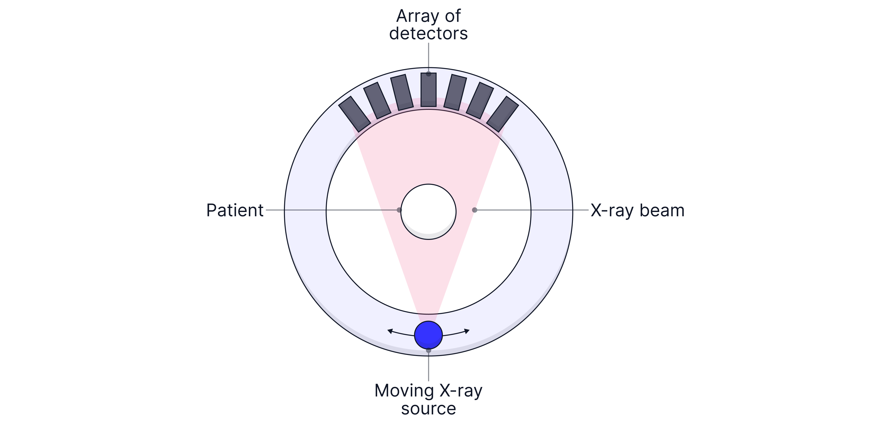
A patient lies in the centre of a ring, and an X-ray tube rotates around the patient, emitting pulses of X-rays. Detectors opposite the source measure transmitted intensity from many different angles. A computer uses these measurements to reconstruct cross-sectional (slice) images of the body. The slices can be combined to produce 3D images of internal structures. The CT detectors contain a scintillator and photodiodes.
Advantages over other imaging techniques:
High-quality images
Non-invasive
Better contrast than ultrasound and can image calcium, unlike MRI
Full cross-sectional area shown
Disadvantages over other imaging techniques:
Exposed to ionising radiation that damages DNA.
Expensive
The patient must remain still, which can cause discomfort
Less contrast between similar soft tissues
Worked Example:
In the past, head injuries were assessed using standard X-ray imaging, but today CT scanning is commonly used instead.
Explain why CT scans have largely replaced simple X-ray images for the assessment of head injuries, while standard X-ray procedures are still appropriate for diagnosing other types of injuries.
In your answer, you should:
outline the fundamental working principles of a CT scanner
compare the benefits of CT scans with simple X-ray images when examining head injuries
justify why simple X-ray imaging remains suitable for assessing other injuries
Answer:
Patient lies in centre of ring with an X-ray tube on one side of the ring with a detector array on the opposite side.
A narrow pulse of X-rays passes through the head detected by the detector array.
The signals from the detector are passed to a computer.
The X-ray tube and detectors are moved about the patient’s head so X-rays are directed through the patient’s head at different angles.
These individual images are compiled and a 2D image of that slice of the head is produced by the computer
For advantages of CT scan:
Better quality image of tissue boundaries inside skull
Can identify bleeding inside skull
For simple X-rays:
Cheaper and easier
Allows simple fractures to be identified
Lower radiation dose received by the patient than a CT scan
Practice Questions
A beam of X-rays passes through of bone. The linear attenuation coefficient of bone is . Calculate the fraction of the original X-ray intensity that is absorbed.
-> Check out Hannah's video explanation for more help.
Answer:
A patient is suspected of having a fractured bone and an X-ray image is required. The image could be produced using one of the following detection systems:
Photographic film
A flat-panel digital detector
Fluoroscopic image intensifier with an intensifying screen.
Identify which detection system would be the most appropriate for this situation and justify your choice.
Explain why the other two systems are less appropriate for imaging a broken bone.
-> Check out Hannah's video explanation for more help.
Answer:
No need to use a fluoroscopic image intensifier as the image is not moving - just want a still image to analyse
A digital detector is more sensitive than photographic film so a lower radiation dose can be used
Digital images are easy to transfer
Using a digital detector is quicker than using photographic film as it takes time to darken the film.