Ultrasound Imaging
Brook Edgar & Hannah Shuter
Teachers


Contents
Explainer Video
Piezoelectric Effect
Ultrasound is a longitudinal pressure wave with frequencies above the range of human hearing ().
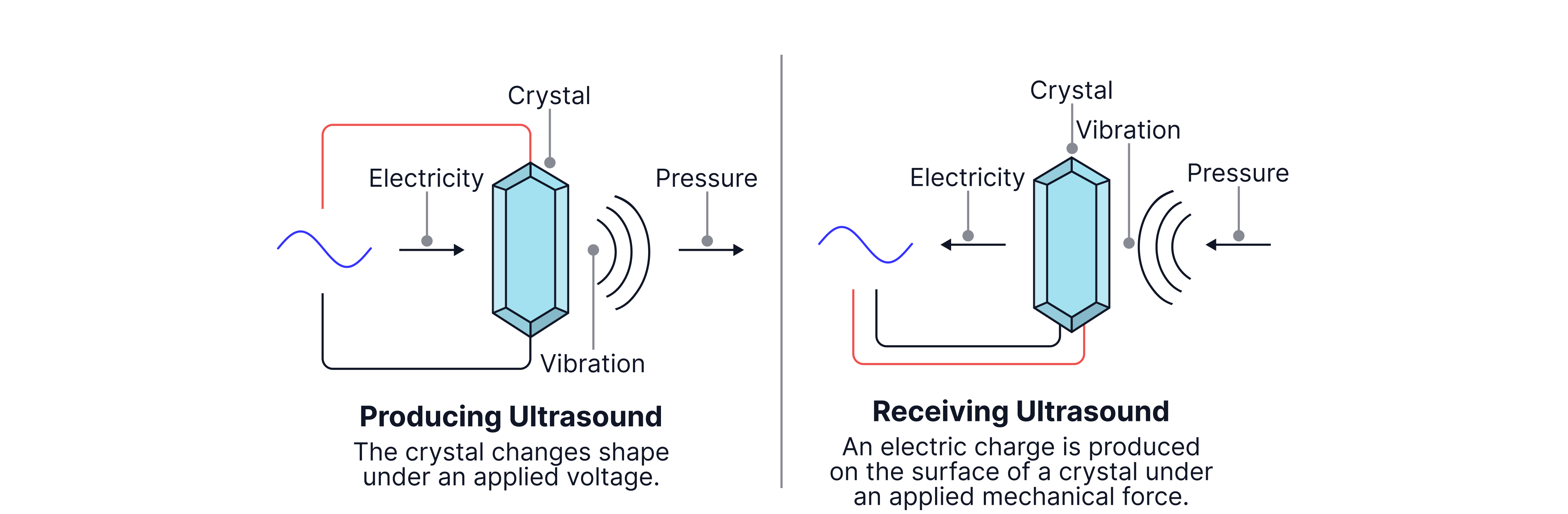
Ultrasounds use high-frequency sound waves to produce images of the body, as waves are reflected at boundaries between different media and detected. Ultrasound waves are both produced and detected by a transducer - a machine that contains a crystal that can be subjected to the piezoelectric effect.
The piezoelectric effect converts mechanical vibrations into electrical energy and vice versa. The transducer is moved across the body and an alternating pd is applied to the crystals. This causes them to mechanically deform, expanding and contracting (vibrating). If the pd is applied at the same frequency as the crystals' natural frequency, it causes them to resonate (vibrate at their maximum amplitude). As the crystals expand and contract, a pressure wave is generated, and an ultrasonic wave is emitted.
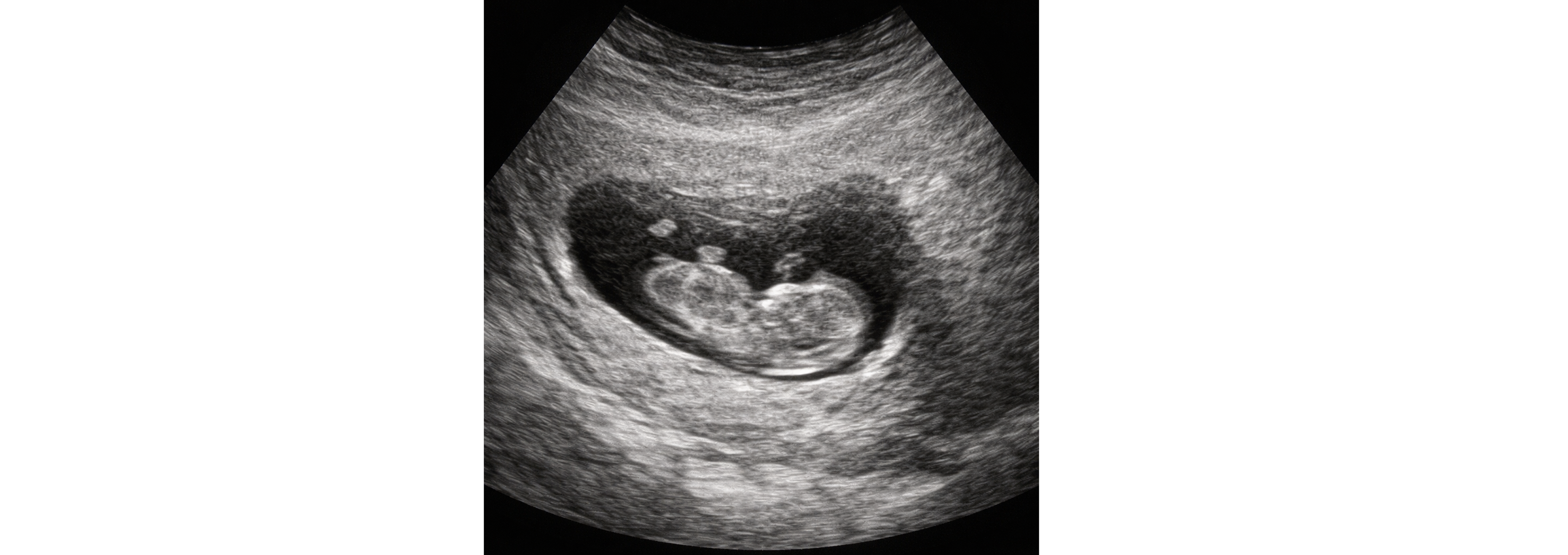
When the reflected ultrasonic wave strikes the crystals, they deform, generating an alternating pd. This pd is then amplified and displayed on a cathode-ray tube (CRT)/oscilloscope, like the image above of an unborn baby. The resolution is poor due to the long wavelengths of ultrasound, (~), however ultrasound is widely used as an imaging technique in obstetrics (a branch of medicine that deals with pregnancy and childbirth) as it is non-invasive and non-ionising, meaning that it will not harm the mother or unborn baby. It produces images in real time, allowing movement to be seen, and is a cheap, quick, and portable imaging technique.
The image below shows the different parts of a transducer.

To get clear, distinct echoes, short pulses are used. To achieve a short pulse, the crystal must stop vibrating quickly, which is achieved using damping. A backing material is placed behind the crystals in the transducer to do this. This improves resolution because the emitted and received signals do not overlap, allowing the transducer to act as both an emitter and a receiver.
Worked Example:
1. Image resolution is an important factor to consider when imaging the body. Explain what resolution is.
2. Estimate the resolution of an ultrasound scan used to image a person's liver, that uses a frequency of . The speed of sound in the liver is .
Answer:
1. Resolution is the ability to distinguish between points that are close together. The smaller the wavelength used, the smaller the structures that can be seen.
2.
Acoustic Impedence
Acoustic impedance is a measure of how much a material resists the passage of sound waves. The acoustic impedance of air, .
Formula:
The greater the difference in acoustic impedance between the two media, the more reflection occurs, such as at a bone-soft tissue boundary or an air-soft tissue boundary.
The air-soft tissue boundary has a large difference in acoustic impedance so a lot of reflection occurs here. This is why, when ultrasound scans are performed, a gel/coupling medium is used between the transducer and the skin to minimise reflections here. This is also why structures behind the lungs are difficult to image: the air-filled lungs reflect rather than transmit most of the ultrasound waves. We cannot image structures behind bone due to bone's high acoustic impedance, so, for example, the brain cannot be imaged by ultrasound.
Intensity of reflection coefficient, , calculates the proportion of the incident wave that is reflected,
, the intensity of reflection coefficient = 0, very little to no reflection occurs.
Worked example:
Backing material is used in a transducer. Explain why.
Ultrasound is transmitted from muscle to bone. Calculate the percentage that is transmitted using the data below:
acoustic impedance of bone =
density of muscle =
speed of ultrasound in muscle =
Answer:
1. Backing material is used to dampen the vibrations of the crystals after the pulse is transmitted so that the transducer can act both as an emitter and a receiver, so that the pulses remain separate.
First, we calculate the acoustic impedance of muscle,
Now we calculate the proportion that is reflected,
If is reflected, then is transmitted.
Remember: Sound travels fastest in solids because the particles are tightly bound and transmit vibrations quickly. Although solids resist motion more (high impedance), this does not slow the wave, because speed depends on stiffness, not on resistance to passage.
A-Scans and B-Scans
A and B-scans both use ultrasound waves to image the inside of a patient, but have different uses. An A-scan is used when only a one-dimensional measurement is required, for example, when the surface of interest lies along the same line, such as measuring the distance between the front and back of a baby's head or the length of the eye. The ultrasound echoes are displayed as peaks on a graph. A B-scan is used to produce a two-dimensional image of more complex structures. The ultrasound echoes are displayed as dots whose brightness depends on their strength.
A-scan (Amplitude Scan)
A transducer emits a short ultrasound pulse (lasting a few microseconds) into the body. At the same time, a spot on the cathode ray oscilloscope (CRO) begins moving horizontally, so the x-axis represents time. When a reflected wave hits the transducer, the crystals vibrate, producing an electrical signal. This signal appears as a peak on the oscilloscope's y-axis and returns to baseline when the echo stops. 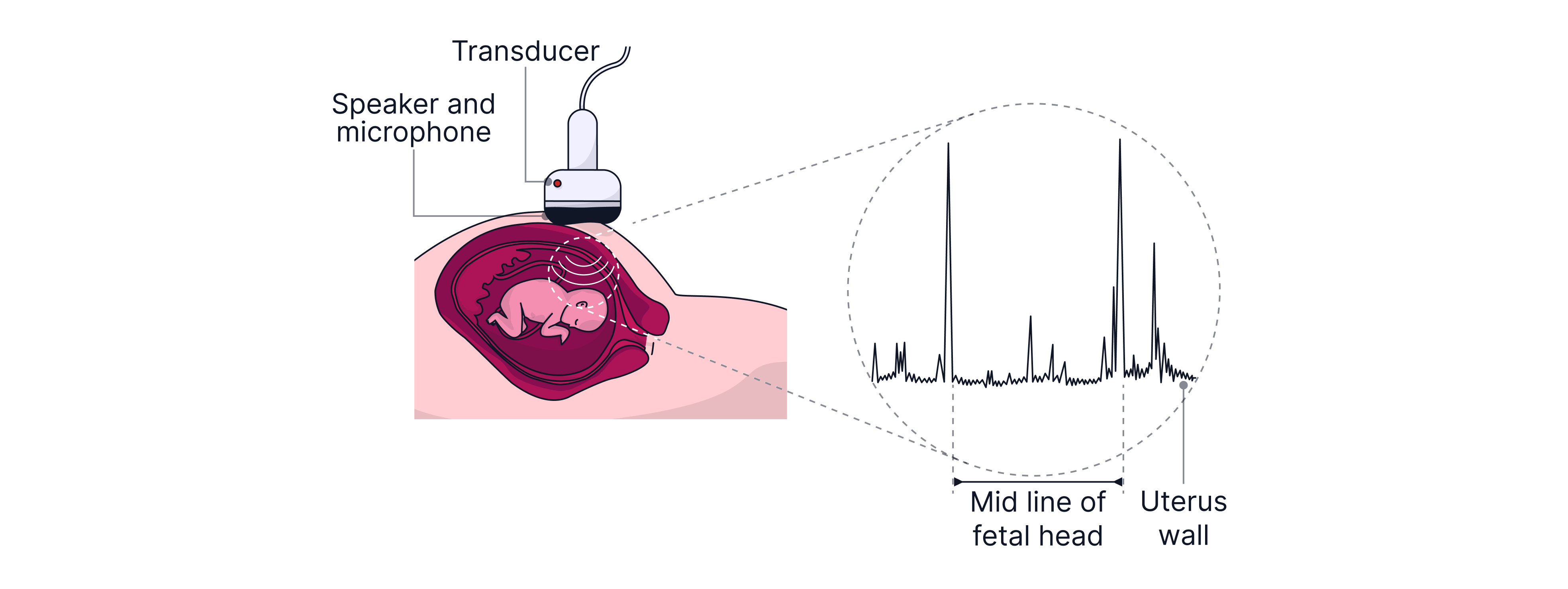 The peak's displacement from the beginning indicates the time it took for the wave to be emitted and reflected back. If the wave's velocity is known, the distance to the boundary can be calculated - be careful to use half the total time, as this is the time to reach the boundary and return! Different peaks represent reflections from different boundaries at different depths/distances inside the body. This is useful, for example, if you wanted to calculate the diameter of a foetus's head to estimate how long until birth, or to check the distance between the lens and the back of the eye.
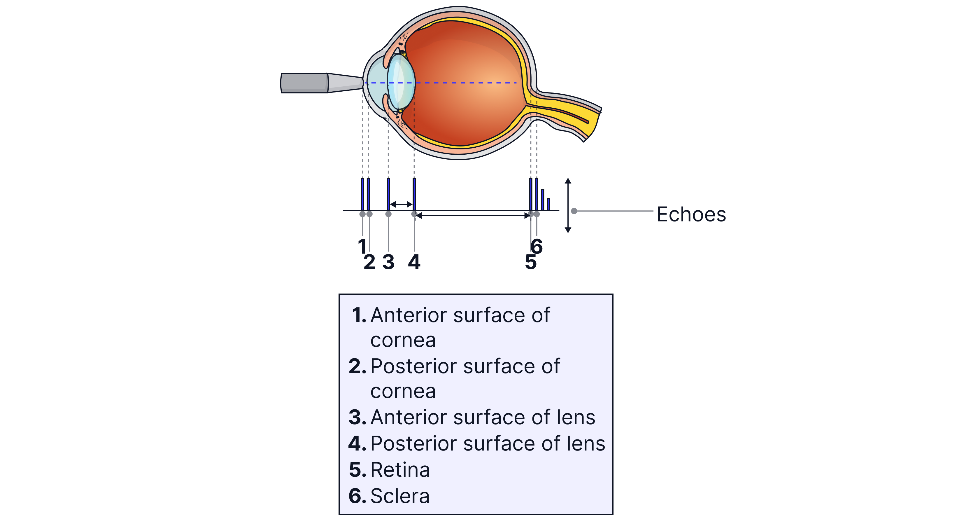
Echo pulses need to be amplified, and signals from echoes returning from greater depths in the body need to be amplified more. Ultrasound waves are attenuated (intensity/energy drops due to absorption/scattering) as they travel through tissue, so echoes from deeper structures are weaker and require more amplification than echoes from shallower structures. This is achieved using a swept-grain amplifier connected to the oscilloscope's time-base setting. The higher the acoustic impedance, , of the medium the more the wave is attenuated.
B-scan (Brightness Scan)
B-scans produce images of body sections. The echoes are used to control the brightness of a spot displayed on a screen. The transducer is moved across the body, and potentiometers (variable resistors that produce a voltage proportional to their position or angle) convert linear motion along the patient into x-axis voltages and angular rotations/tilts of the transducer (since the transducer can only receive a signal when it is normal to the reflected beam) into y-axis voltages. These coordinates plot a point on the oscilloscope screen to indicate the transducer position. As the transducer must be tilted, the operator must be skilful and experienced to ensure that contact with the gel is maintained and that no air gap forms.
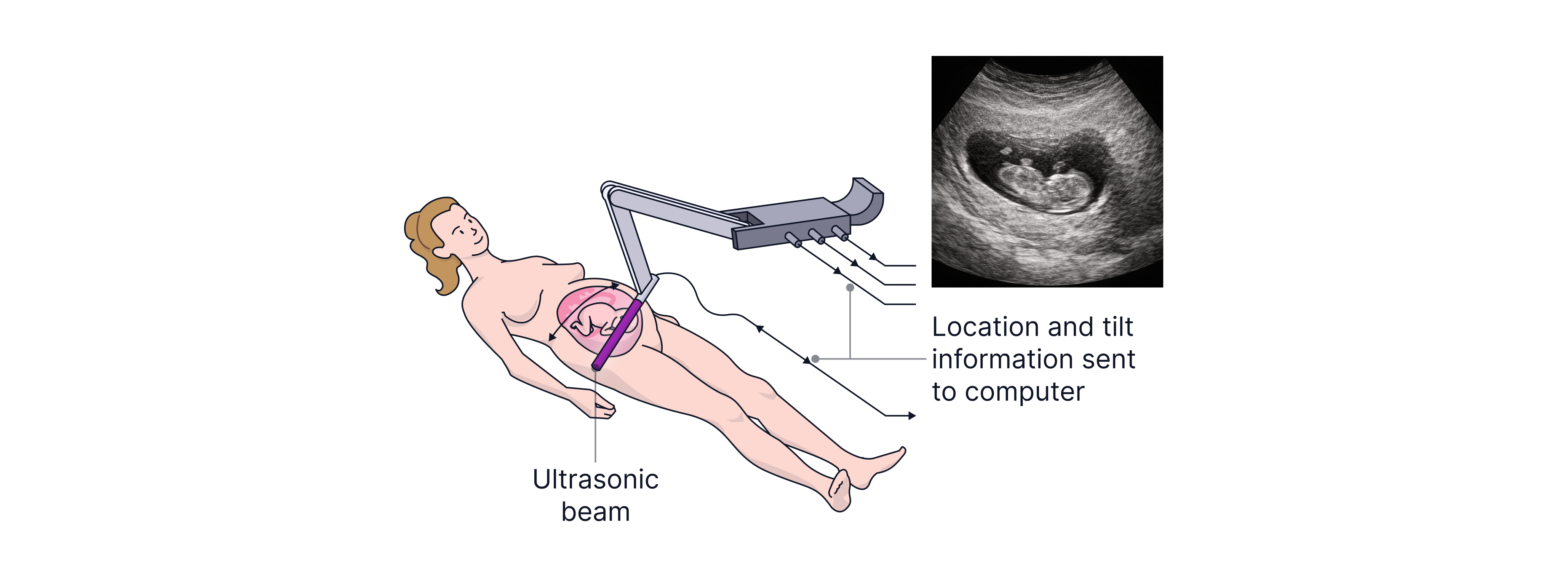
A B-scan is used to determine the position of the baby's head midline, and an A-scan is then used to measure the head diameter.
B -scans are also important to determine the position of the placenta before birth, as if it is covering the cervix, a C-section will need to be carried out.
Worked Example:
A coupling gel needs to be applied to the patient's skin when getting an ultrasound scan. Explain why.
Answer:
A coupling gel is needed because of the large difference in acoustic impedance between the skin and air. Without one, a very small percentage of waves would be transmitted, as most would be reflected. The gel used needs to have an acoustic impedance close to that of the skin to maximise transmission.
Practice Questions
The image is of an ultrasound transducer. Explain how it works.
-> Check out Brook's video explanation for more help.
Answer :
An alternating potential difference is applied across the crystals. The frequency of the alternating potential difference is equal to the resonant frequency of the crystal
This causes the crystals to expand and contract
This creates pressure waves that are emitted with a frequency above
To record ultrasound, the pressure waves are received by the crystals, causing them to expand and contract, inducing an alternating pd
Using short pulses of alternating pd allows the transducer to act as a transmitter and a receiver. When the potential difference is removed, a backing material is used to damp and stop the vibrations of the crystals so that they can receive the reflected ultrasound.
The image shows the A-scan of an ultasound of a patients abdomen.

A) State two reasons why the amplitude varies.
B) Ultrasound travels at in soft tissue. Estimate the distance between the front of the patient's abdomen and their spinal cord.
-> Check out Brook's video explanation for more help.
A) The amplitude of pulses is attenuated as it penetrates deeper (reduction in intensity due to absorption).
It also depends on the acoustic impedance of the two materials at the interface - the proportions of ultrasound which are reflected or transmitted
*remember you divide the final distance by two as it is a reflection