ECG, MRI and Fibre optics
Brook Edgar & Hannah Shuter
Teachers


Brook Edgar
Teacher
Hannah Shuter
Teacher
Explainer Video
ECG
An ECG (electrocardiogram) is a recording of the heart's electrical activity produced by measuring changes in potential difference over time and displaying the information on an oscilloscope screen.
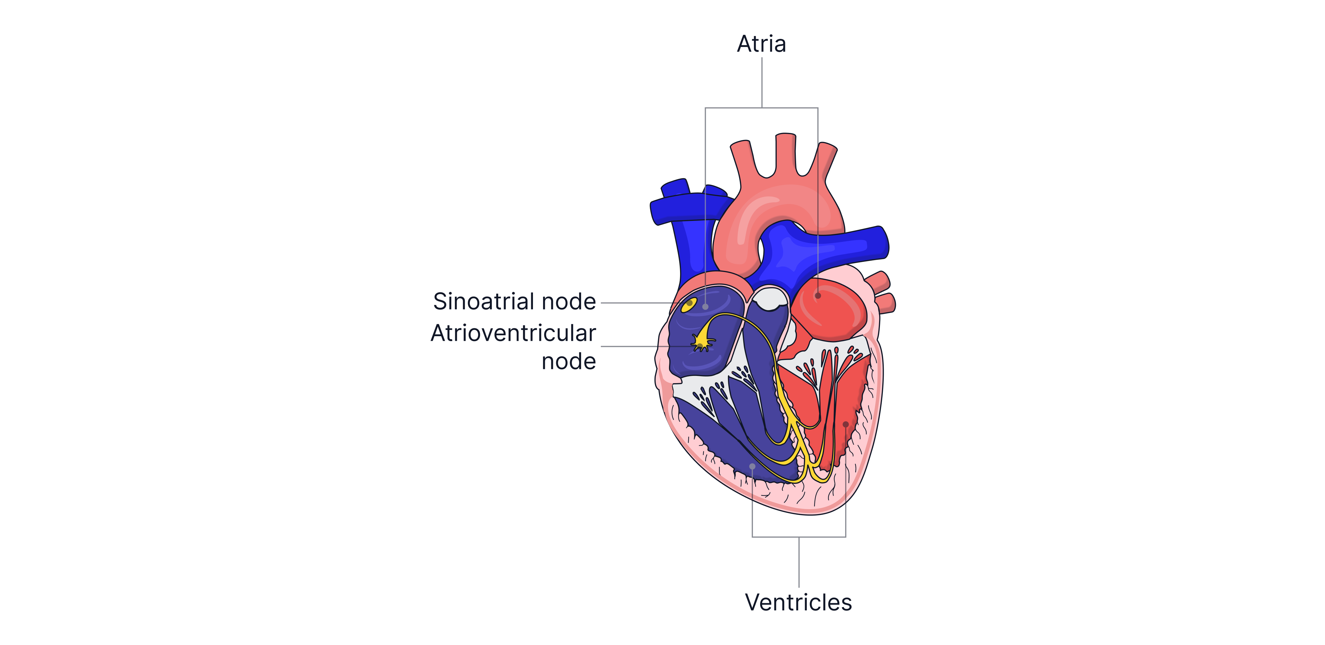
Electrical signals in the heart control its contraction, pumping blood from the heart to the lungs and around the body. The heart is a double pump, and each side of the heart is separated into two chambers called the atria and ventricles. The atria and ventricles are separated by a valve to prevent the backflow of blood. Electrical signals are generated in the atria by the sinoatrial node (SA node) and then spread across both atria, causing them to contract. This causes the ventricles to fill with blood. There is a short, deliberate delay to allow time for the blood to move before the atrioventricular node (AV node) fires, causing the ventricles to contract and move blood out of the heart. The AV node is delayed as its cells conduct more slowly than other cardiac cells.
To produce the ECG, metal plate electrodes are attached firmly in pairs to different parts of the body (can be anywhere arteries are close to the surface) to measure the heart's electrical activity. This can be done by measuring the the voltage difference on the skin, because electrical impulses spread throughout the body from the heart via electrolytes in the blood and interstitial fluid (fluid between cells in tissues). The body is rubbed with an abrasive material to remove dead skin and hair to improve the electrode connection. A conducting electrode gel is also applied to the skin, containing electrolytes to enhance electrical contact between the skin and metal electrodes. As the skin is nonconductive, any air gap between the electrode and the skin would reduce the connection. The metal electrodes are designed to avoid reacting with chemicals in the skin.
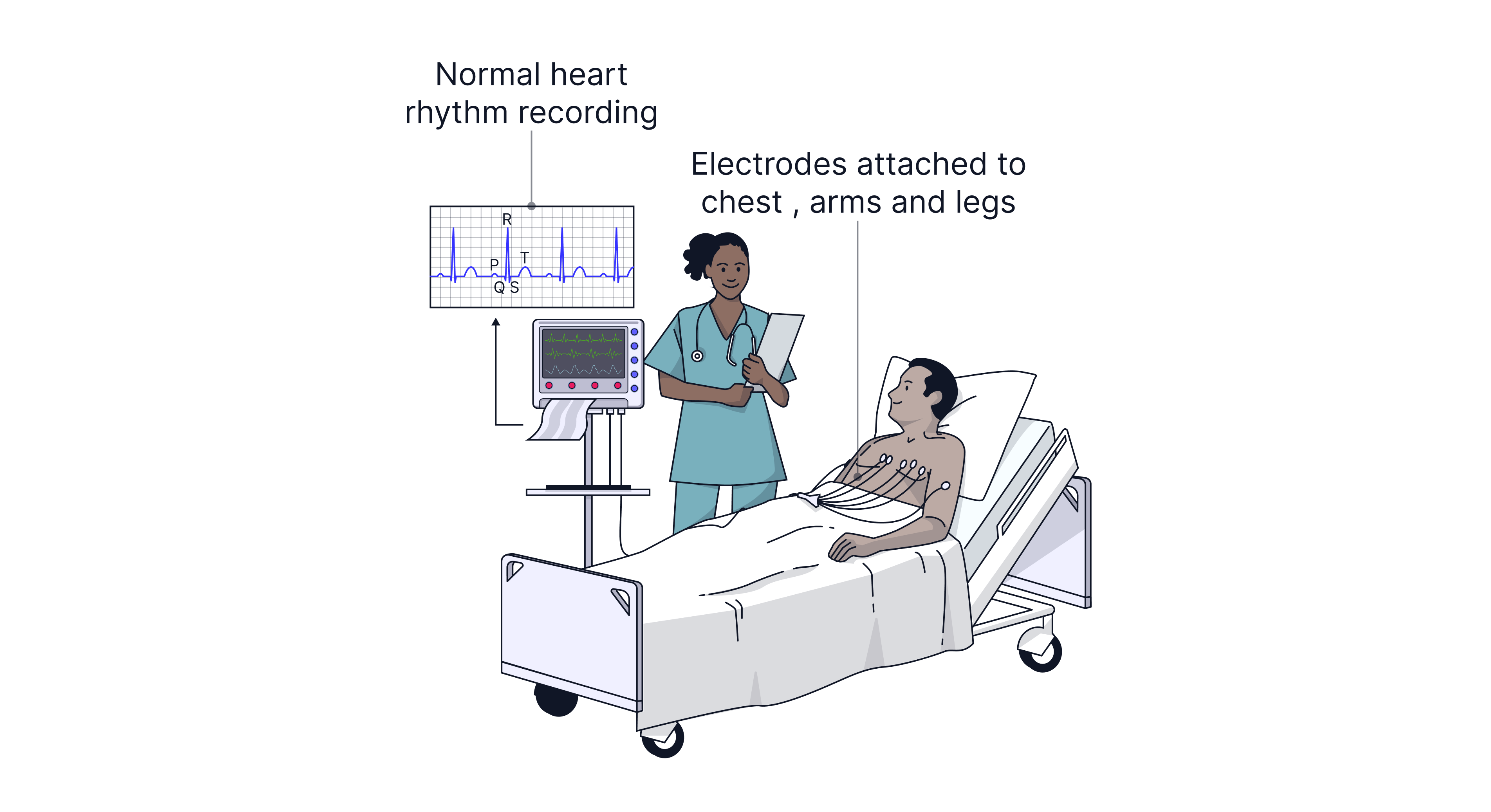
The patient needs to be as relaxed as possible and remain still, as twitching/muscle contractions can be picked up on the ECG. This unwanted electrical noise is biological in origin however electrical equipment can also produce unwanted noise. The human body acts like one plate of a capacitor and nearby cables as the other. The alternating electric field in the cables induces a charge on the body (capacitive coupling), and the alternating magnetic field induces a pd on the body (Faraday's law) and in the wires (inductive coupling). The electrocardiograph and leads are hence shielded to reduce this unwanted electrical noise.
The detected signal from the heart is amplified because the electrical impulses are weak, but this also amplifies electrical noise. The electrodes, therefore, measure the potential difference between each pair to minimise noise, since unwanted biological and electrical signals are of the same order of magnitude across all connections. The amplifier must be high-gain to amplify the tiny signals detected at the skin and low-noise, meaning it adds very little to no unwanted signal of its own. It is shielded so the reading is not affected, and it has a high input resistance so the signal received is not distorted, since high resistance will not draw any current from the electrodes themselves.
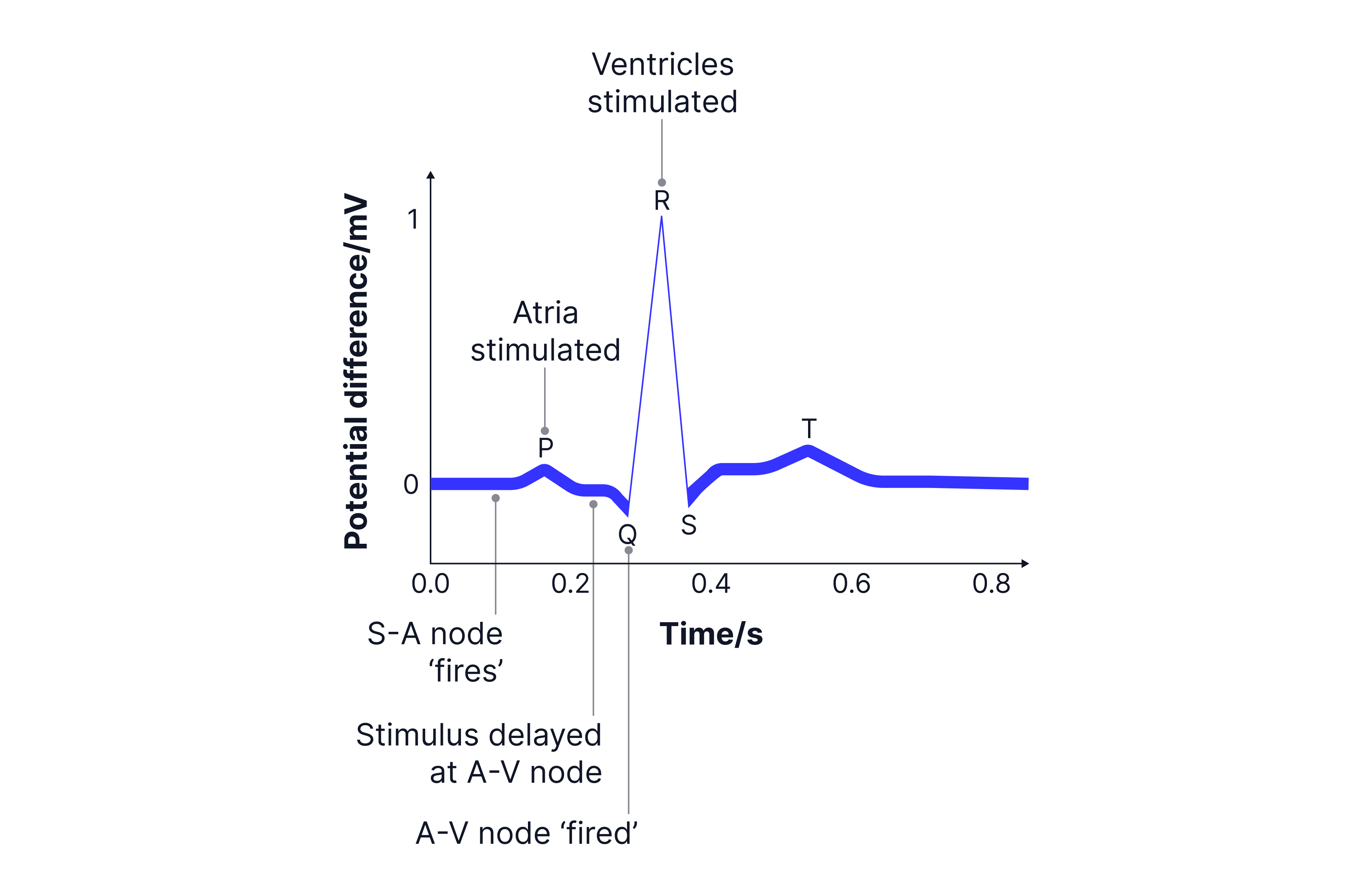
The ECG waveform shows a P wave when the electrical signal is first produced in the SA node, causing the atria to contract.
The QRS peak occurs around seconds later when the signal leaves the AV node, causing the ventricles to contract. The height of the peak corresponds to the amplitude of the pd detected at the skin -> .
The T wave happens around another seconds later and occurs when the ventricles relax in preparation for the next heartbeat (the relaxation of the atria is not shown/detected as the signal is hidden by the large spike caused when the ventricles contract).
To find the pulse rate, you measure the time between two R points from separate heartbeats (the period of the wave). To find the pulse rate per minute, calculate how many waves pass in seconds.
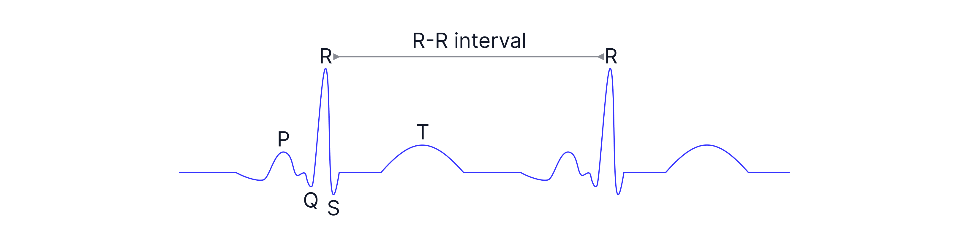
We can use ECGs to see the differences when a person exercises, as the period would decrease, so their pulse rate would increase, and we can also use it to detect if there are any abnormalities in the heart’s rhythm.
Worked Example :
Describe the procedure used to ensure that a good ECG trace is obtained from a patient.
Explain:
How the skin is prepared, and a property of the gel used
How unwanted signals are avoided
Properties of the amplifier
Answer:
To obtain a good ECG, ensure good electrical contact between the electrodes and the skin by using conducting gel.
You also need to ensure the electrodes are attached firmly to reduce noise by preventing them from moving.
You will want to rub the skin with an abrasive material to remove dead skin cells and hair, reducing contact resistance.
The electrodes should be nonreactive with chemicals in the skin, and the gel used should be nonirritating to the skin.
To further reduce noise, you will want the patient to remain still and relaxed and to use shielding around the amplifier and leads to reduce electrical interference from nearby AC equipment.
The amplifier should have high gain to amplify the low signals received, low noise, and a large input impedance (high resistance) to avoid drawing current from the electrodes.
MRI
MRI is magnetic resonance imaging. The machine uses a strong magnetic field to change the orientation of protons (hydrogen nuclei) in the human body. The person is inside a ring of detectors that measure radiation emitted by hydrogen nuclei, generating 3D and 2D cross-sectional images of the body.
The body is mostly water, so it contains many hydrogen nuclei. Protons have a quantum property called spin, which makes them behave like tiny bar magnets. Spin is not motion through space, but it is a built-in quantum property, like mass and charge, that behaves mathematically like angular momentum. Because the proton carries a charge, its spin gives it a magnetic moment (moving charges -> current -> magnetic field). The strong, static (uniform) magnetic field created by superconducting magnets aligns the hydrogen nuclei parallel to it. However, the protons do not align perfectly but wobble around the magnetic field lines; this is known as precession (similar to how a spinning top wobbles under gravity).

Smaller electromagnets, known as gradient coils, are used to produce overlapping magnetic fields across the body, resulting in a gradient in the magnetic field strength in the x, y, and z directions. Therefore, as protons in different parts of the body experience different magnetic field strengths, they will have different precessional frequencies. A proton with the same precession frequency as the radio-frequency (RF) signal emitted by the MRI machine will become excited and change its spin (alignment).

When the radio pulses are stopped, the hydrogen nuclei de-excite and realign with the magnetic field. Their spins change so they emit RF signals at their precession frequencies. These EM waves are detected and processed to build an image on the computer, since their exact positions are known. It is the gradient in the static magnetic field that causes different radio-wave frequencies to be emitted at different locations along the body. If there were no gradient, all protons would precess at the same frequency, so no spatial information would be given.
The time it takes for the nuclei to realign after the radio wave is turned off is called the relaxation time, and different tissue types have different relaxation times, leading to brightness differences in images due to varying signal strengths. You are not required to know about relaxation times, but it helps to understand how the image is built up.
As only magnetic fields and radio waves are used, there is no ionising radiation so an MRI is not dangerous. There are no harmful effects on the patient or the machine operator. It is a non-invasive (no cutting) imaging technique and produces real-time images. However, if the patient has any metallic implants in their body, such as a pacemaker (a small electronic device that sends electrical impulses to the heart to ensure it beats at a normal rate), an MRI scan cannot be performed. A further disadvantage is that the patient has to remain still for a long time, and the machine's noise can cause discomfort. It is an expensive imaging technique and cannot image calcium and thus bone well.
The MRI machine's ability to distinguish between different tissue types is better than that of CT scans (more on this later), and it has higher resolution than ultrasound and CT scans.
Worked Example:
During an MR brain scan, a patient is exposed to a strong magnetic field and short pulses of radio-frequency electromagnetic waves. Explain the principles of the MR scanner. Use the image to help you. 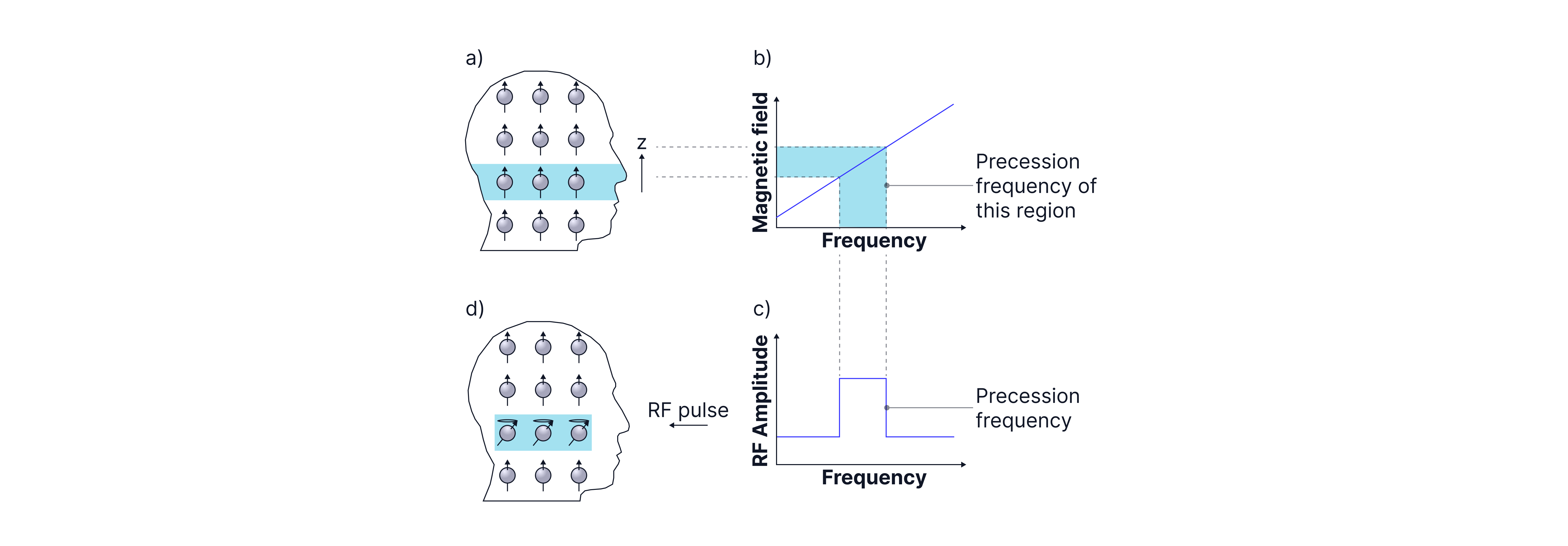
Answer:
A strong magnetic field is generated using superconducting magnets, which align the protons with the field (as shown in part a of the diagram).
Electromagnets are used to produce a gradient in the static magnetic field, so hydrogen nuclei in different regions of the body have different precessional frequencies (part b).
Radio pulses are applied at specific frequencies, exciting hydrogen nuclei at specific locations, changing their spin (part c).
When they de-excite, they emit radio signals (part d). The signals are detected and passed to a computer, and an image of the body is built up.
Fibre Optics
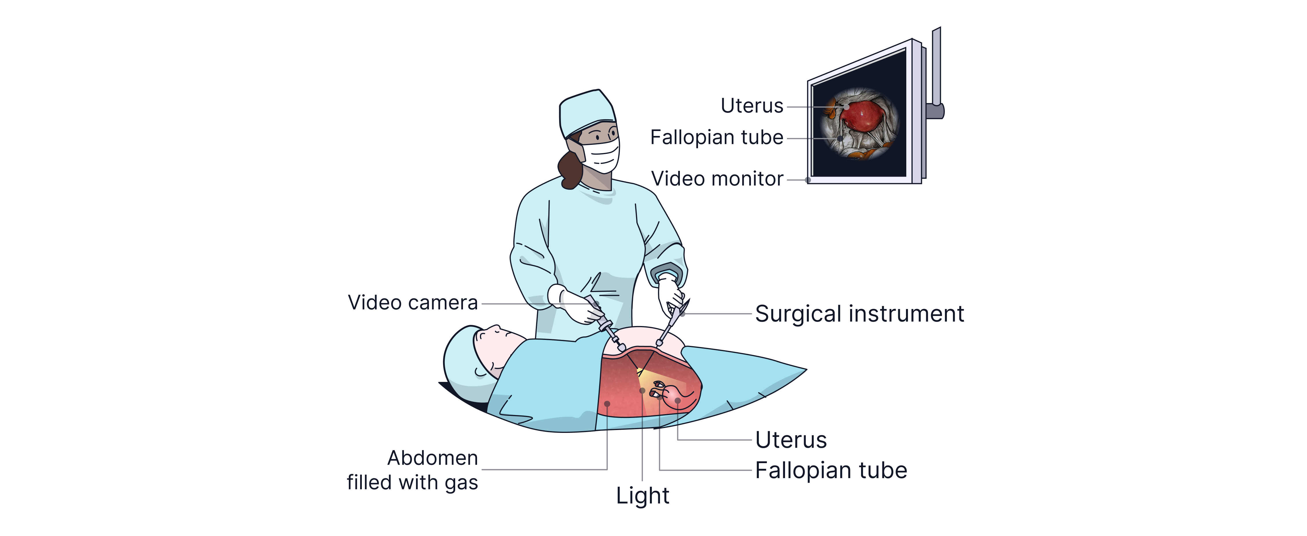
In medical practice, a small incision can be made to examine the inside of the body. This is done by inserting one bundle of optic fibres to illuminate the area and another to transmit the image. These bundles are known as endoscopes. Surgical tools, such as cutters or cauterising devices, can be passed through an endoscope to carry out operations without making a large incision. This technique, known as keyhole surgery only uses only small incisions, reducing the risk of infection, minimising damage to healthy tissue, shortening recovery time, and lowering healthcare costs.
Optical fibres use total internal reflection (TIR). An optical fibre must be transparent so that light can pass through the core without being absorbed or scattered, allowing the signal to travel long distances. Inside each cable is a bundle of optical fibres, but their walls cannot touch as this would cause light to leak from one to another. To prevent this, the fibres are cladded in a glass layer (which has a lower refractive index than the core) to isolate each fibre, preventing signal degradation (reduction in quality/strength of the signal) from light escaping and protecting the fibres. The cables can be curved, but if too curved, the angle of incidence at the core-cladding boundary is likely to fall below the critical angle, causing signal degradation as light escapes.
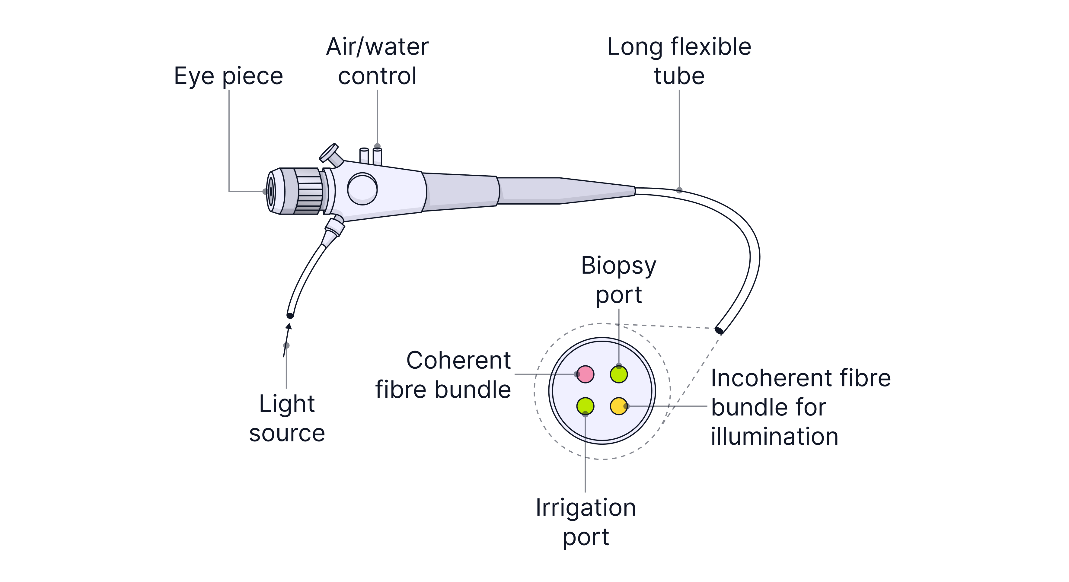
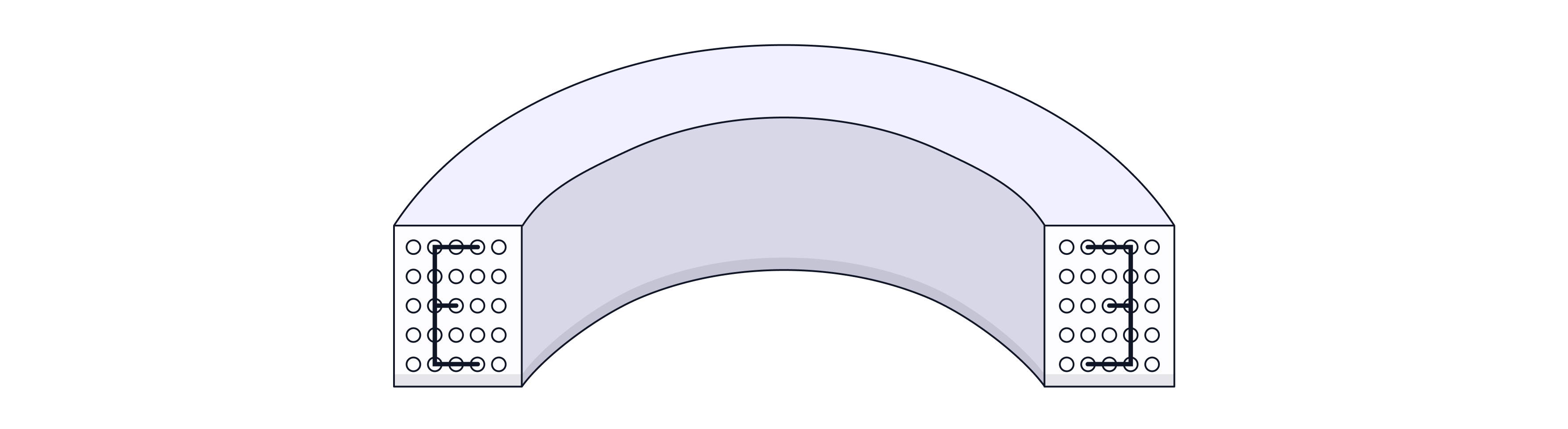
Bundles of optic fibres that carry images need to be coherent, meaning the relative arrangement of the optic fibres is kept the same along the bundle, as each part of the fibre transmits a small part of the image. Image quality improves when fibres are tightly packed with very small diameters, thereby increasing resolution and allowing more detail to be seen. The image can be magnified if the fibre diameter increases along its length toward the eyepiece lens.
Bundles of fibres that carry light into the body to illuminate it do not need to be coherent. It does not matter where each fibre is positioned; they are arranged randomly and do not require cladding, which makes them cheaper to produce. They are known as incoherent bundles.
Worked Example:
Explain why methods to reduce modal and material dispersion in optic fibres are not required in an endoscope.
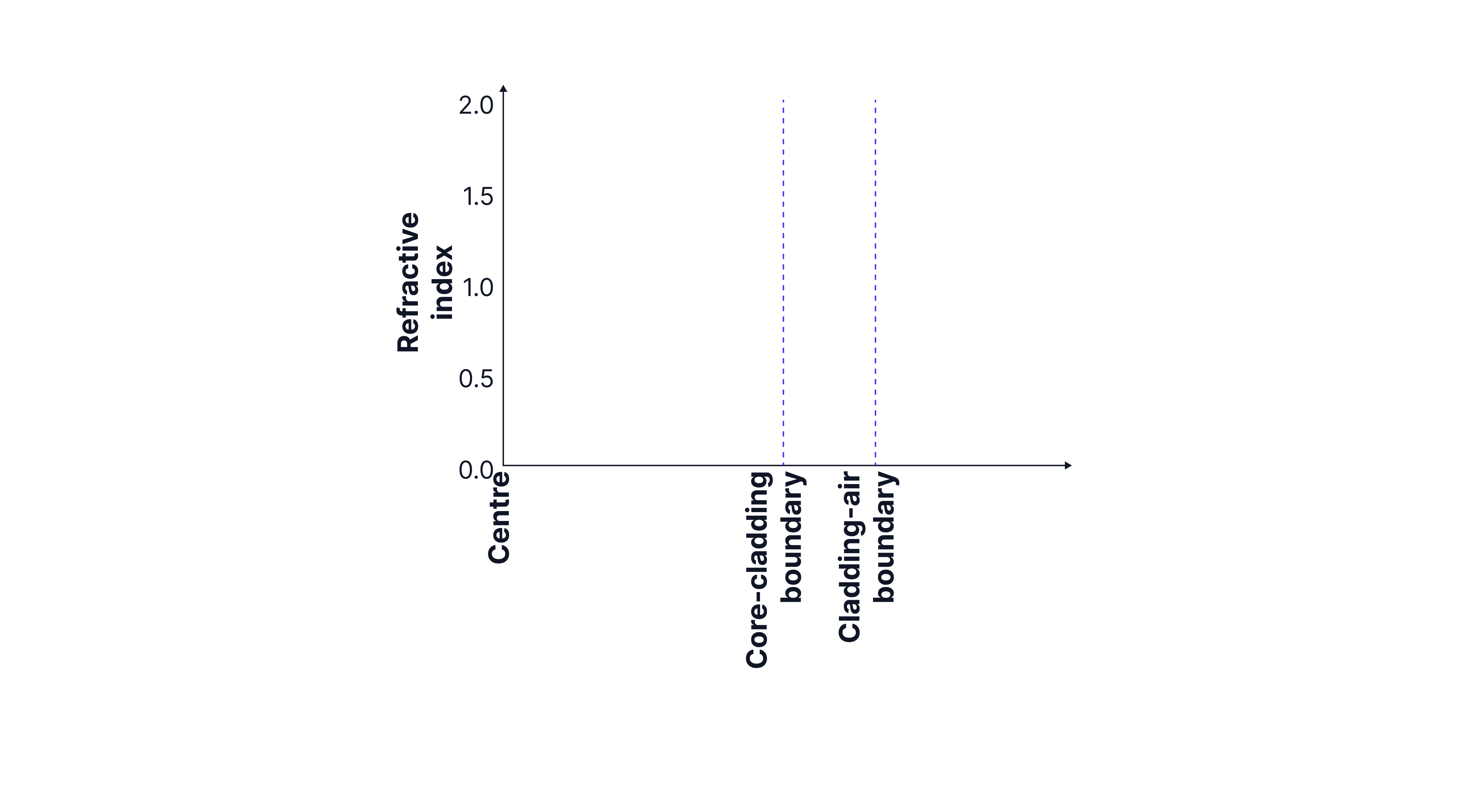
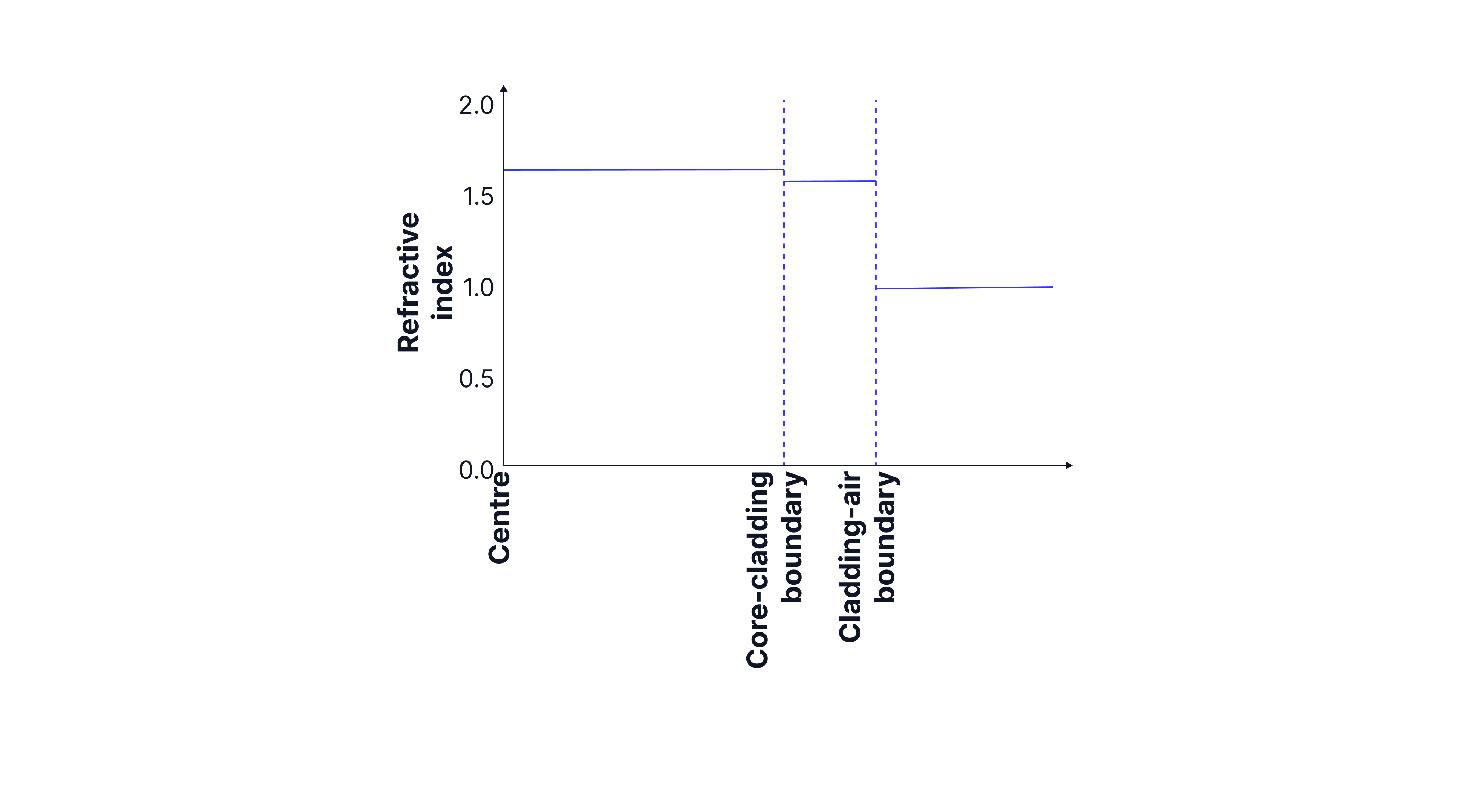
An optic fibre has a critical angle of degrees at the core cladding boundary and is placed in air. The refractive index of the core is . Complete the graph below showing how the refractive index varies with distance from the centre of the core.
Answer:
When transmitting data over long distances, to ensure that all the light arrives at the same time, monochromatic light is used to reduce pulse broadening. This is known as material dispersion. Endoscopes transmit light only over short distances, and if monochromatic light were used, images would be in only one colour, so little detail would be visible.
Narrow fibres are used to reduce modal dispersion caused by different angles of incidence. This could be useful, as more fibres could be fitted into the bundle, improving image resolution, but it is expensive and unnecessary when the endoscope is used for illumination only. Also, the short distances involved mean that there is not much path difference.
Repeaters are not needed as the endoscope is short, so the signal does not need to be amplified.
We know the refractive index of the core -> 1.6, and that of air -> 1.
We can calculate the cladding's refractive index using the critical angle at the core-cladding boundary.
Practice Questions
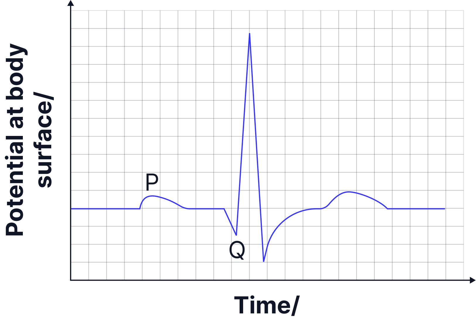
Label the scales in the ECG trace of a healthy person.
State the electrical event that occurred and the physical change that resulted from points P and Q on the graph above.
-> Check out Brook's video explanation for more help
Answer:
x-axis in mV, peak at 1 mV. y-axis in seconds.
Position P: Sino atrial node fires, atria contracts.
Position Q: Ventricular node fires, ventricles contract.
An endoscope has a coherent and non-coherent fibre bundles.
State the use of each bundle.
The core of each fibre has a refractive index of and the cladding has a refractive index of
Calculate the critical angle in degrees.
-> Check out Brook's video explanation for more help.
Answer:
Coherent bundles are used to transmit images out of the body, non-coherent bundles are used to transfer light into body/to illuminate.
State the purpose of the magnetic field in a magnetic resonance scanner.
-> Check out Brook's video explanation for more help
Answer:
Align spins of hydrogen nuclei/protons.
OR
Causes hydrogen nuclei to precess around the magnetic field/in one direction.