Animal Cells
Laura Armstrong & Joe Wolfensohn
Teachers


Recall Questions
This topic builds on GCSE knowledge of basic animal cell structure, the names and functions of key organelles, and the differences between animal and plant cells.
Name four parts of an animal cell.
Nucleus, mitochondria, ribosomes, cell membrane.
State the function of the mitochondria.
Mitochondria are the site of aerobic respiration.
Give two ways in which an animal cell differs from a plant cell.
Animal cells do not have a cell wall, chloroplasts or a large permanent vacuole.
Topic Explainer Videos
Check out these videos that explains Animal Cells, then read the study notes. Once you’ve gone through them, don’t forget to try the practice questions!
What is an animal cell?
Animal cells are eukaryotic cells - they contain a true nucleus enclosed by a nuclear envelope and a range of membrane-bound organelles in the cytoplasm. This distinguishes them from prokaryotic cells such as bacteria.
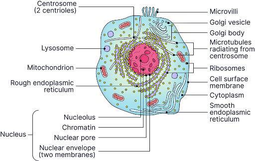
The structure of an animal cell.
The Nucleus
-
Surrounded by the nuclear envelope, a double membrane containing nuclear pores.
-
Nuclear pores allow the passage of substances such as mRNA out of the nucleus.
-
Contains chromatin (DNA wound around histone proteins), which condenses into chromosomes during cell division.
-
Contains the nucleolus, where ribosomal RNA (rRNA) is synthesised.
-
The nucleus controls the activities of the cell by directing protein synthesis.
Mitochondria
-
Surrounded by a double membrane (outer membrane and inner membrane).
-
The inner membrane is folded into structures called cristae, which increase the surface area for the reactions of aerobic respiration.
-
The fluid-filled interior is called the matrix; it contains enzymes, mitochondrial DNA, and 70S ribosomes.
-
Function: site of aerobic respiration and ATP synthesis.
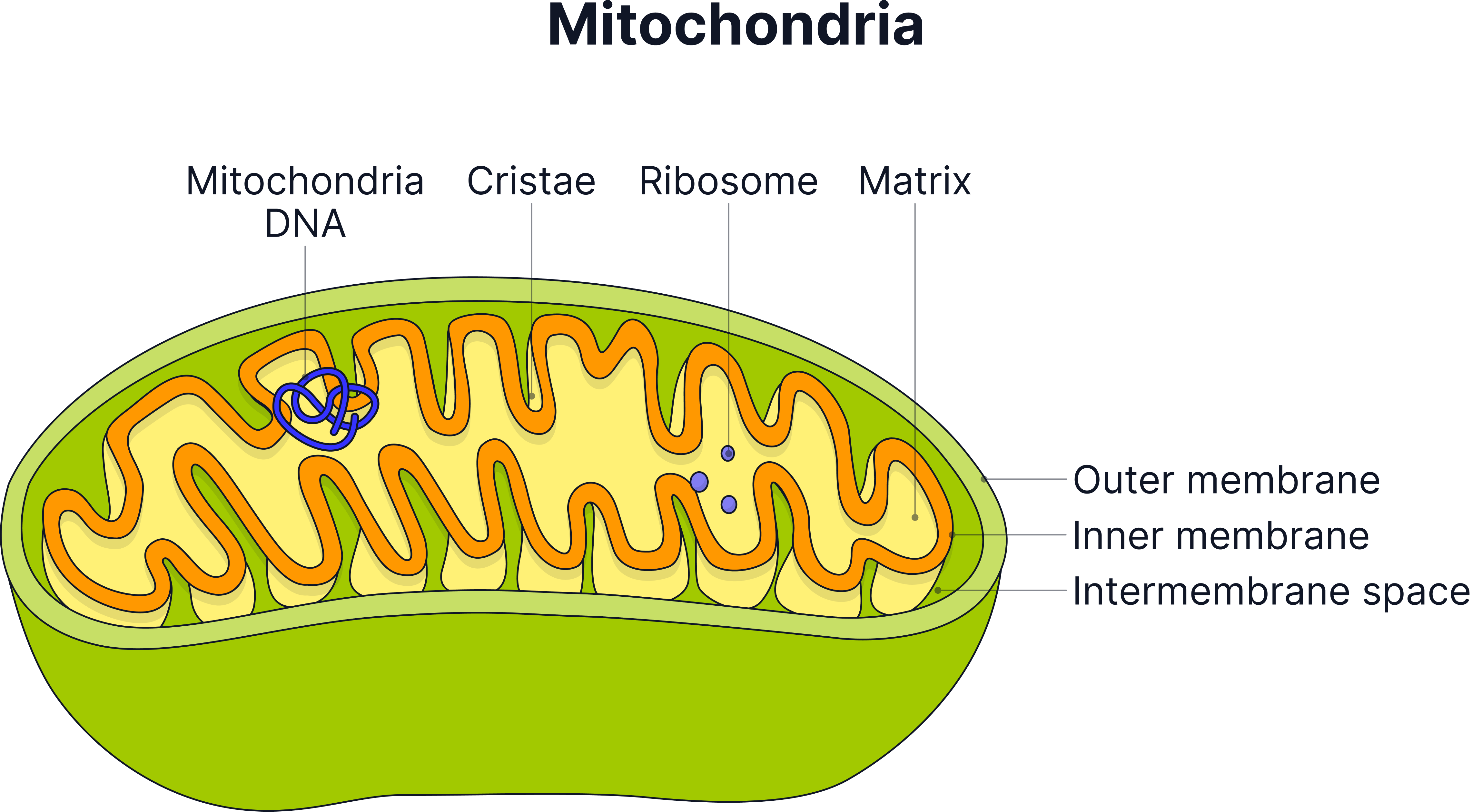
Diagram of the structure of a mitochondrion.
Ribosomes
-
Very small organelles, not membrane-bound.
-
Animal cells contain 80S ribosomes (made up of a large 60S subunit and a small 40S subunit).
-
The subunits are made from protein and rRNA.
-
Function: site of protein synthesis (translation of mRNA).
-
Note: mitochondria contain their own 70S ribosomes, which are smaller than the cytoplasmic 80S type.
Endoplasmic reticulum (ER)
-
A network of membrane-bound channels extending from the nuclear envelope.
-
Rough endoplasmic reticulum (RER): has ribosomes on its surface; involved in the synthesis and transport of proteins.
-
Smooth endoplasmic reticulum (SER): no ribosomes; involved in the synthesis and transport of lipids.
Golgi apparatus
-
A stack of flattened, membrane-bound sacs called cisternae.
-
Receives vesicles from the ER, modifies and packages the proteins or lipids, then buds off vesicles for transport or secretion.
-
Produces lysosomes.
Lysosomes
-
Small, spherical, membrane-bound organelles.
-
Contain hydrolytic (digestive) enzymes.
-
Function: break down worn-out organelles, digest material taken in by phagocytosis, and cause controlled cell death (apoptosis).
Cell surface membrane
-
Surrounds the cell and controls the movement of substances in and out.
-
Made of a phospholipid bilayer with embedded proteins (fluid mosaic model).
The cytoskeleton
The cytoskeleton is a dynamic network of protein fibres that extends throughout the cytoplasm of eukaryotic cells. It maintains cell shape, allows cell movement, enables the transport of organelles and vesicles within the cell, and forms the structural basis of cilia and flagella.
There are three main components:
A) Microfilaments
-
Made of the protein actin, arranged as two strands twisted together.
-
Functions: maintain and change cell shape; involved in cytokinesis (division of the cytoplasm and cell surface membrane) and enable movement in cells such as phagocytes.
B) Microtubules
-
Hollow cylinders made of the protein tubulin.
-
Larger than microfilaments.
-
Functions: form the spindle fibres that separate chromosomes during mitosis and meiosis; act as tracks along which motor proteins move organelles and vesicles; form the core structural element of cilia and flagella.
C) Intermediate filaments
-
Made of a variety of fibrous proteins (for example, keratin in epithelial cells).
-
Intermediate in size between microfilaments (~7 nm) and microtubules (~25 nm).
-
Functions: provide mechanical strength and resistance to stress; anchor organelles (particularly the nucleus) in position within the cell.
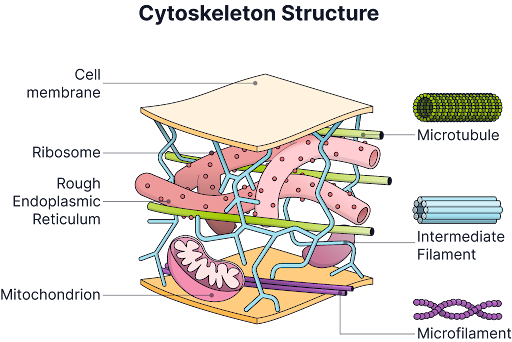
The structure of the cytoskeleton.
Centrioles
-
Pairs of short cylindrical structures found in animal cells, located in a region called the centrosome.
-
During cell division, centrioles organise the mitotic spindle by directing microtubule assembly.
Cilia
Cilia are short, hair-like projections from the cell surface, found on many animal cells. They are present in large numbers and beat in a coordinated, rhythmic pattern.
Structure of a cilium:
-
Each cilium is bound by an extension of the cell surface membrane.
-
The core of the cilium contains nine pairs of microtubules arranged in a ring around two central single microtubules. This is known as the 9+2 arrangement.
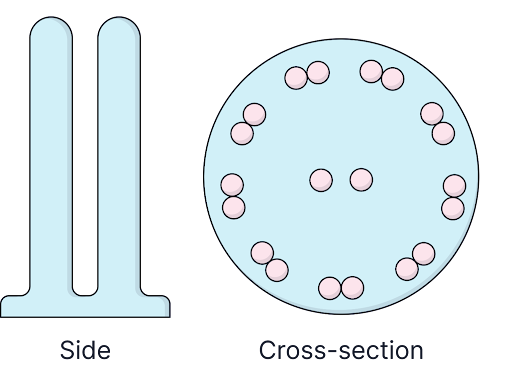
The 9+2 formation of cilia and flagella
Functions of cilia:
-
Moving substances across a surface: the coordinated beating of cilia on the epithelial cells lining the trachea (windpipe) moves the mucus layer - along with trapped particles and microorganisms - upward towards the throat, clearing the airways.
-
Moving the cell: some single-celled eukaryotes use cilia for locomotion, though this is less relevant to animal cell biology at A-level.
Flagella in eukaryotes
Eukaryotic flagella share the same fundamental 9+2 microtubule structure as cilia. The key differences are:
-
Flagella are much longer than cilia and are usually present singly or in small numbers (rather than the large numbers of cilia on a cell surface).
-
Flagella produce a wave-like or whip-like motion rather than the symmetrical back-and-forth beat of cilia.
-
The best-studied example in human biology is the flagellum of a sperm cell, which drives the swimming motion of the sperm towards the egg.
Important distinction - eukaryotic vs prokaryotic flagella:
-
Prokaryotic flagella (e.g. in bacteria) are structurally entirely different. They are made of the protein flagellin, are solid (not hollow microtubules), and rotate like a propeller rather than bending.
-
Eukaryotic flagella are membrane-bound, hollow, and use the 9+2 microtubule arrangement.
Key Terms
-
Eukaryotic cell: a cell that contains a true, membrane-bound nucleus.
-
Cell surface membrane: the phospholipid bilayer that forms the outer boundary of the cell.
Exam Tips
-
Use the correct term for the outer boundary of the cell. Examiners have noted that many candidates write "plasma membrane," "cell membrane," or "bilayer." The preferred OCR A term is cell surface membrane. Using a vague term risks losing the mark.
-
When asked to compare animal and plant cells, always make a comparative statement. For example, write "animal cells do not have a cell wall, whereas plant cells do" rather than simply "no cell wall." A non-comparative statement often does not gain the mark.
Practice Questions
A patient is found to have a mutation that prevents the Golgi apparatus from producing functioning vesicles. Which of the following effects would most likely result from this mutation?
A) The cell would be unable to carry out aerobic respiration.
B) Proteins produced at the rough endoplasmic reticulum would not be modified or secreted.
C) The nucleus would be unable to direct protein synthesis.
D) The cell surface membrane would no longer be selectively permeable.
Answer: B
Rationale:
The Golgi apparatus receives vesicles from the RER, modifies proteins, and packages them into vesicles for secretion. If vesicle production fails, this pathway is blocked. A is wrong; aerobic respiration occurs in mitochondria and is unrelated to the Golgi. C is wrong; the nucleus directs transcription independently of the Golgi. D is wrong; the cell surface membrane's permeability depends on its protein and lipid composition, not directly on Golgi vesicle function.
A student is examining a tissue sample from a mammalian liver.
(i) State two features visible in the image that would confirm the cell is eukaryotic rather than prokaryotic. [2
(ii) The student identifies an organelle with a double membrane and internal folds. Identify this organelle and explain how its internal structure is related to its function. [3]
(iii) The student then observes a small, spherical, membrane-bound organelle that contains electron-dense material. Suggest the identity of this organelle and give one function it could carry out in a liver cell. [2]
Model answer:
(i)
-
The cell contains a membrane-bound nucleus, enclosed by a nuclear envelope
-
Membrane-bound organelles are present, such as mitochondria, endoplasmic reticulum, or the Golgi apparatus
(ii)
-
The organelle is a mitochondrion
-
Its inner membrane is folded into structures called cristae
-
These folds increase the surface area available for the enzymes involved in ATP synthesis / oxidative phosphorylation, making aerobic respiration more efficient
(iiI)
-
The organelle shown is a vesicle
-
It contains enzymes or transport proteins, which it carries to their destination within or outside the cell
More Practice
Try to answer the practice questions from the TikTok videos on your own, then watch the video to see how well you did!