Structure of the Eye
Brook Edgar & Hannah Shuter
Teachers


Contents
Brook Edgar
Teacher
Hannah Shuter
Teacher
Explainer Video
Structure of the Eye
The Sun emits electromagnetic radiation across the entire spectrum, but the human eye evolved to be sensitive only to wavelengths between , known as the visible spectrum. When photons within this range enter the eye, they are detected by photoreceptor cells in the retina and converted into electrical signals. These signals are transmitted to the brain, where they are interpreted as different colours. The perceived brightness of an image depends on the intensity (number of photons) of the light reaching the eye.
The figure below shows an image of the eye. The typical diameter of the eye is around .
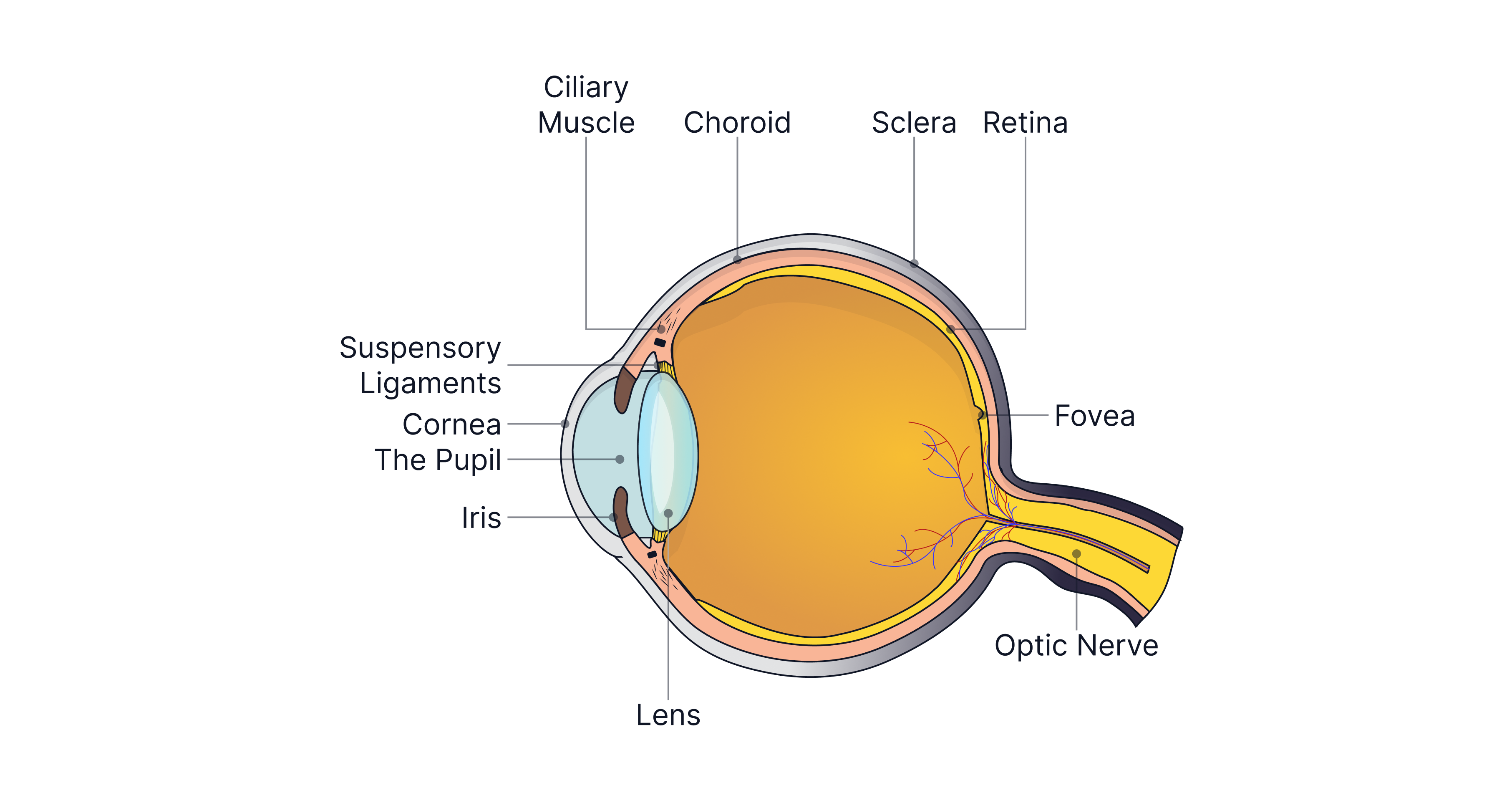
Pupil -> Light enters the eye through the pupil, which acts as the eye's aperture (opening). The amount of light entering the eye is controlled by the iris, the coloured part of the eye. In bright conditions, the iris constricts, making the pupil smaller to reduce the amount of light reaching the retina and protect it. In dim conditions, the iris dilates, enlarging the pupil to allow more light to enter. You can observe this effect by looking in a mirror after spending time in a dark room and then suddenly switching on the lights. The pupil also becomes smaller when viewing nearby objects. A smaller pupil blocks light rays passing through the outer regions of the eye's lens, where they are refracted more strongly than rays passing close to the principal axis. This reduces spherical aberration, allowing a sharper image to form on the retina.
Retina -> At the back of the eye, where photoreceptors (light-sensitive cells) are located, called rods and cones. Rods function in low-light conditions, and because several rods are connected to a single nerve fibre, they provide simple light perception and detail, yielding greyscale images. The fovea (also called the yellow spot) is a small region of the retina directly opposite the pupil. It contains only cones, which are responsible for colour vision. Cones are smaller than rods, and each cone is connected by a single nerve fibre to the brain, providing good spatial resolution, allowing fine detail to be seen. The density of cones decreases, and the density of rods increases as you move further away from the fovea.
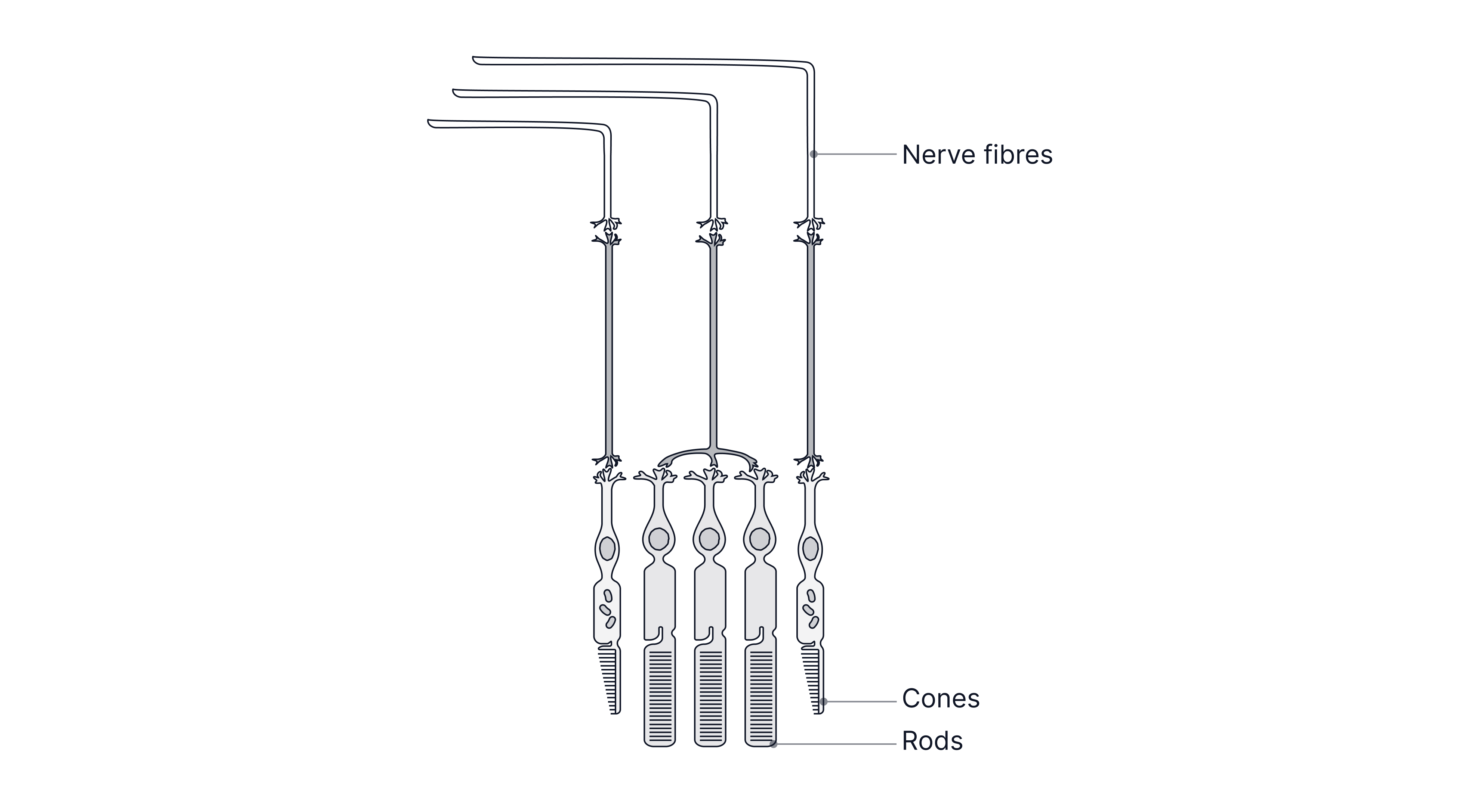 Blind spot -> The part of the eye that is not sensitive to light, because it contains no photoreceptor cells (rods or cones). It is the point where all the nerve fibres from the retina converge to form the optic nerve, which carries visual information to the brain.
Cornea -> A protective layer covering the front of the eye, where most refraction occurs. It is the transparent section of the sclera, the tough protective layer of the eye, that is opaque at the back to prevent light reflections, which would otherwise cause blurred images. The sclera is lined by a layer of tissue called the choroid, which supplies the eye with oxygen and glucose via a network of blood vessels. The spherical shape of the eye is maintained by the pressure of the transparent fluids inside it.
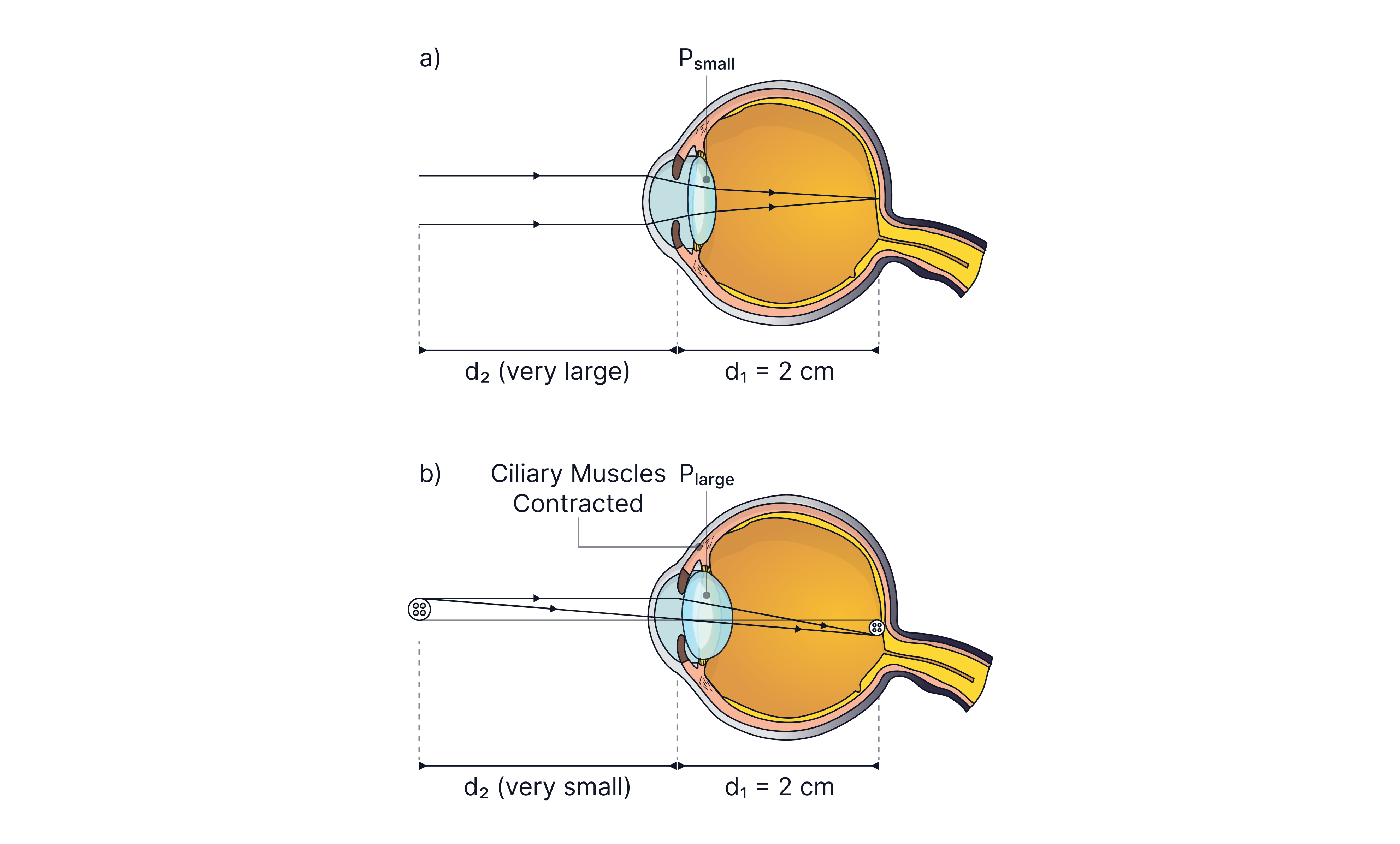
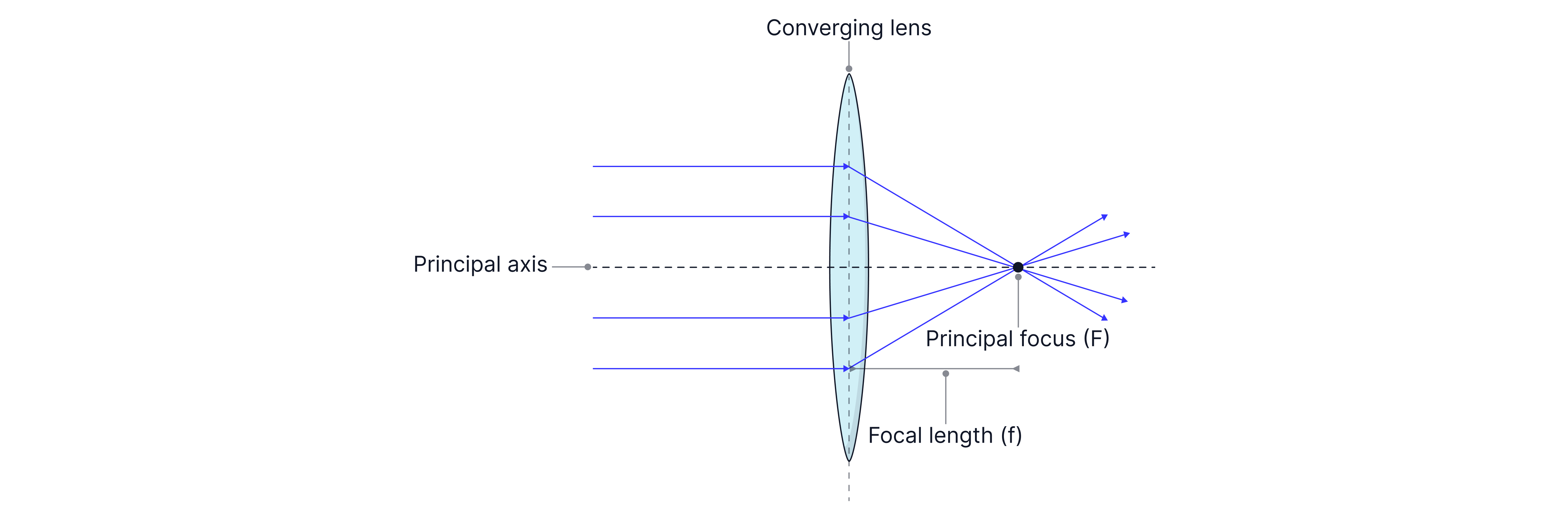
Lens -> Responsible for accommodation, the process by which the eye changes the shape of the lens to focus on objects at different distances. The lens is convex meaning it converges light rays, bringing them to a focus. The lens is held in place by the suspensory ligaments, which are attached to the ciliary muscles. The ciliary muscles control the shape of the lens. Rays of light from distant objects are parallel, so less refraction is required to focus them onto the retina. The ciliary muscles relax, allowing the eye to have a longer focal length between , and a lower refractive power. If an object is closer, a more powerful lens is required to focus the light rays onto the retina. The lens changes shape by contraction of the ciliary muscles, becoming more curved. The near point of the eye - the closest distance at which an object can be focused clearly is .
Worked Example:
State the role of the iris.
What is accommodation?
Describe the changes that occur in the eye from viewing a near object to a distant one.
Describe the changes that occur in the eye from viewing an object in bright light to dim light.
Answer:
The iris controls the intensity of light reaching the retina.
Accommodation is the ability of the eye to change shape by changing the thickness of the lens using the ciliary muscles to focus on objects at different distances.
The eye needs to have a longer focal length when viewing distant objects. The lens needs to be less powerful, which is achieved when the ciliary muscles relax, making the lens less curved/thinner.
In dim light, the cones stop working, and the rods take over. The pupil enlarges/dilates to allow as much light as possible to enter the eye. It takes some time for the eye to adjust.
Reminder: Convex lenses converge light rays, this means that they bring light rays to a focus on the other side of the lens. Lenses use refraction. Refraction is the change in direction of light as it travels from one medium to another, as light changes speed.
Sensitivity of the Eye
Rods and cones are known as photoelectric cells, as they convert light energy into electrical signals (nerve impulses transmitted to the brain).
Rods contain a light-sensitive pigment called rhodopsin or visual purple, which is destroyed (bleached) when light hits it, producing a small emf. An enzyme reverses this process in dim light, but it can take up to , which is why it takes time for the eyes to adapt to the darkness (dark adaptation).
Cones are responsible for colour vision. They contain three types of light-sensitive pigments, each sensitive to one of the primary colours of light: red, green, or blue. The brain perceives other colours by comparing the relative responses of the three types of cones, depending on the intensity of light falling on each type of cone.
Red cones are most sensitive to long wavelengths as red light has the longest wavelength, detecting light in the range .
Green cones are most sensitive to medium wavelengths in the range . The eye is most sensitive to these wavelengths because the Sun emits more photons in the green region of the spectrum due to its temperature, and our eyes have adapted to this.
Blue cones are most sensitive to short wavelengths, from . The eye is least sensitive to blue light, being about one-third as sensitive as it is to green light. Blue light is strongly scattered by the atmosphere, giving the sky its blue colour.

Remember: You need to be able to draw this diagram showing the response curves for the colour cones in the eye. I remember the numbers by knowing the starting wavelengths all differ by .
Spatial Resolution
An image can be resolved if light falls on light-sensitive cells/receptors separated by one unstimulated light-sensitive cell. Therefore, images can be resolved if they are separated by more than two cell diameters on the retina.
You can calculate if objects can be resolved or not by the eye using the equation from GCSE maths, .
Formula:
But when applied to the eye,
You can either know the angular separation of the objects and find out if it covers more than two cell diameters on the retina, or you can be asked to find the diameter of the receptors at the back of the eye, knowing an image is resolved.
For example, if two sources of light subtend an angle of at the naked eye, we can calculate the diameter of the receptor if the eyeball is assumed to be spherical with a diameter of
Worked Example:
State the condition required for two objects to be resolved as separate images on the retina.
Explain how the resolution of an object viewed in very dim white light differs from its resolution when viewed in bright white light.
Answer:
The two images must fall on receptors separated by at least one unstimulated receptor.
In bright light, the cones are used, giving coloured images, and in dim light, the rods are used, giving greyscale images. The resolution is better in bright light, as the cones are smaller and are connected to one nerve cell
Practice Questions
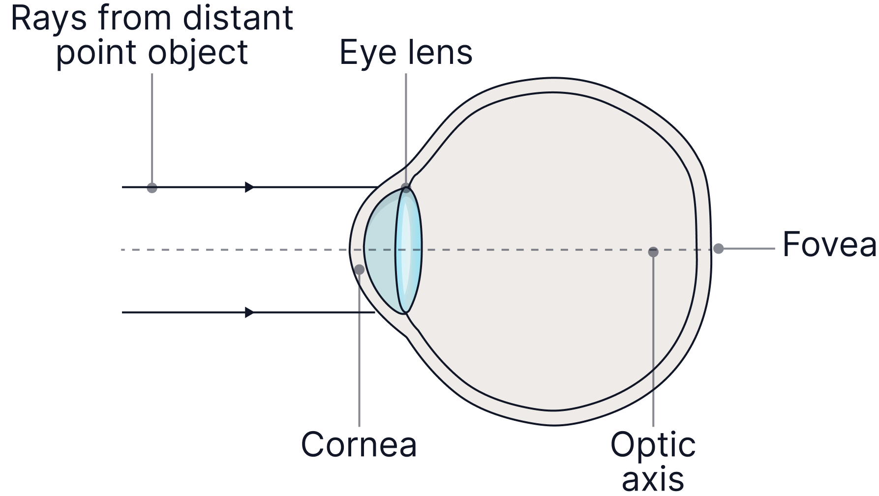
Sketch the path of rays of light from a distant object on the diagram below
Sketch a graph showing the response curves for the three cones.
-> Check out Brook's video explanation for more help.
Answer:
See TT video -> most refraction occurs at the cornea, some at the lens, hitting the fovea at the back of the eye.
The eye can see light in the range . See the TT video showing the graph. Intensity on the y-axis and wavelength in nanometers on the x-axis. Peak intensity of each primary colour at the midpoint of each range.
Determine if two point sources of light, apart, from the eye can be resolved. The diameter of a cone is . The fovea is from the lens.
-> Check out Brook's video explanation for more help.
Answer:
We know the images can be resolved if they are separated by more than two cell diameters on the retina.
We are then going to solve for first, the angular separation of these sources of light.
Now we are going to use their angular separation and the diameter of the eye to solve for , the distance that these two sources of light cover on the retina, and compare this number to the diameter of two cones.
Two cell diameters =
Therefore, they can be resolved.