DNA
Lajoy Tucker
Teacher

Contents
Introduction to DNA
DNA (Deoxyribonucleic Acid) is a large, double-stranded polymer made up of nucleotide monomers. These two complementary strands are arranged in the form of a double helix.
DNA Explainer Video
Structure of DNA
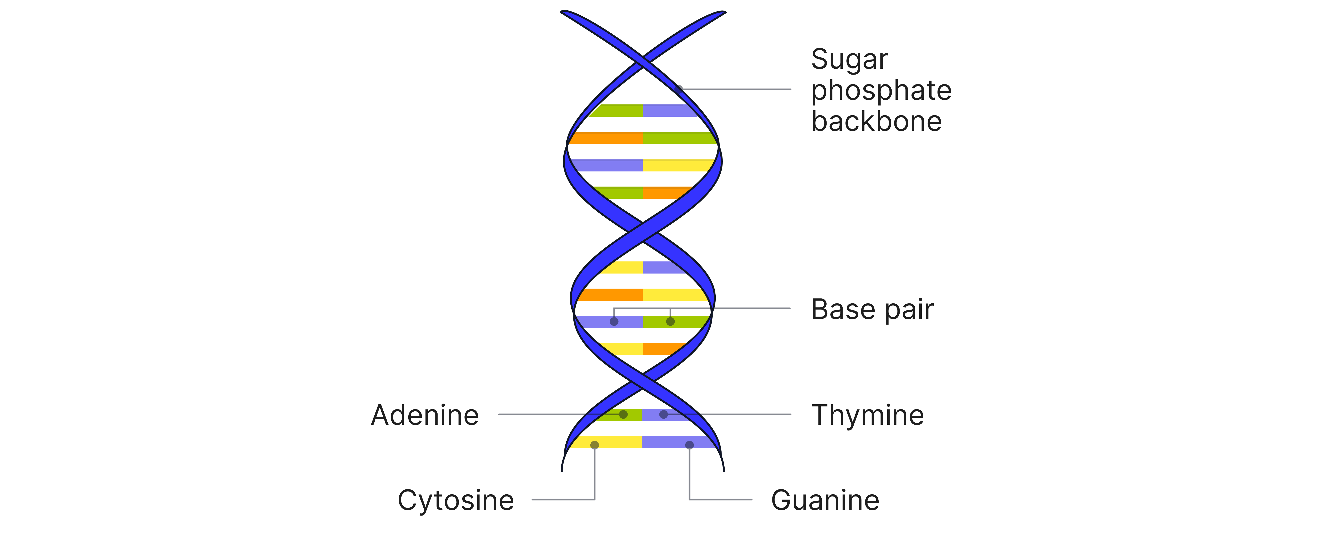
The monomers that make up DNA are nucleotides.
Each nucleotide in DNA consists of:
1. A phosphate group

2. A pentose sugar (2-deoxyribose)

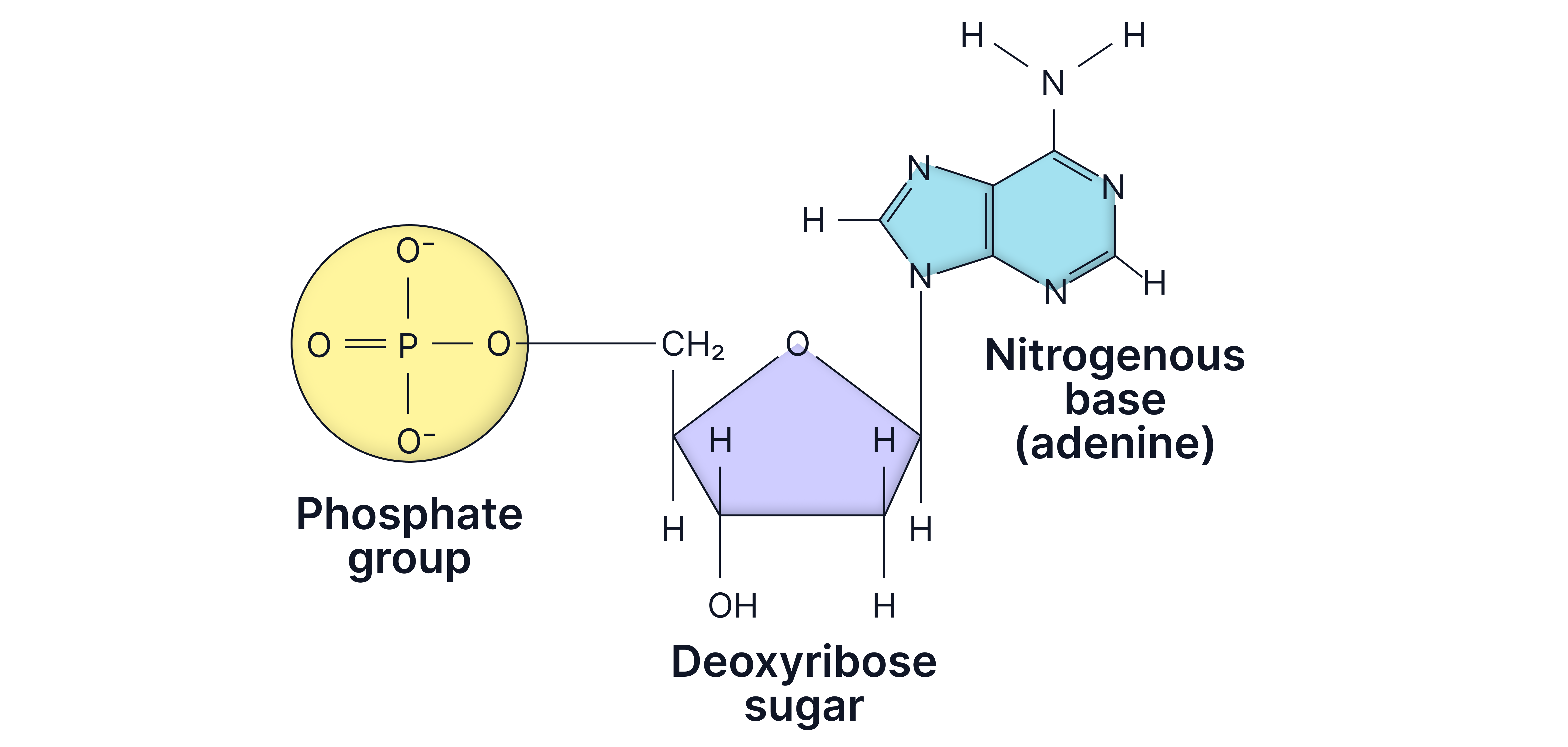
3. A nitrogenous base
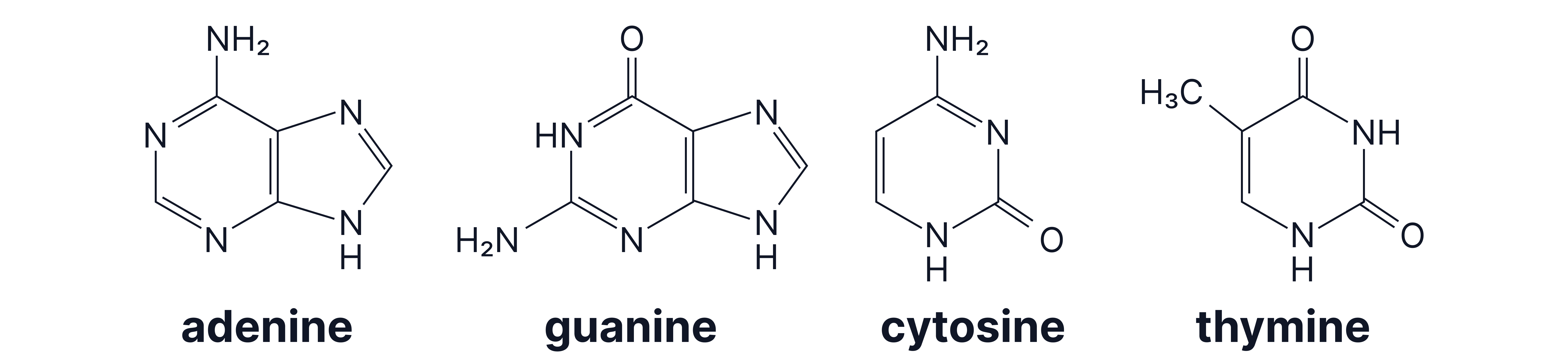
Example nucleotide with adenine as the base
Nucleotides are joined by phosphodiester bonds between the phosphate group of one and the sugar of the next.
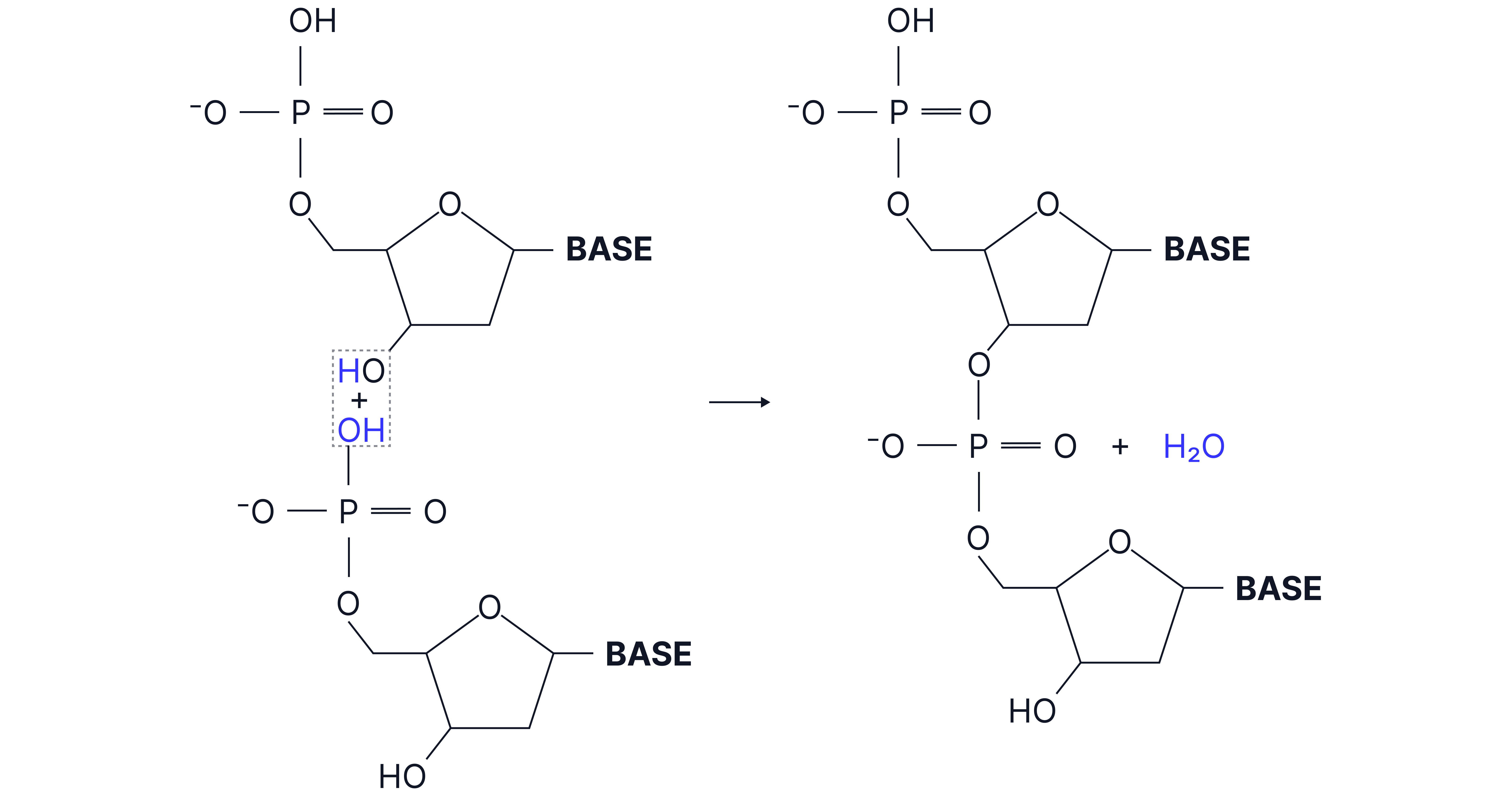
This results in a long strand of repeating nucleotides linked by covalent bonds in a sugar-phosphate-sugar-phosphate polymer chain.
Base-pairing
The double-stranded nature of DNA arises from bases on antiparallel polynucleotide strands pairing up. These base pairs are held together by hydrogen bonds.
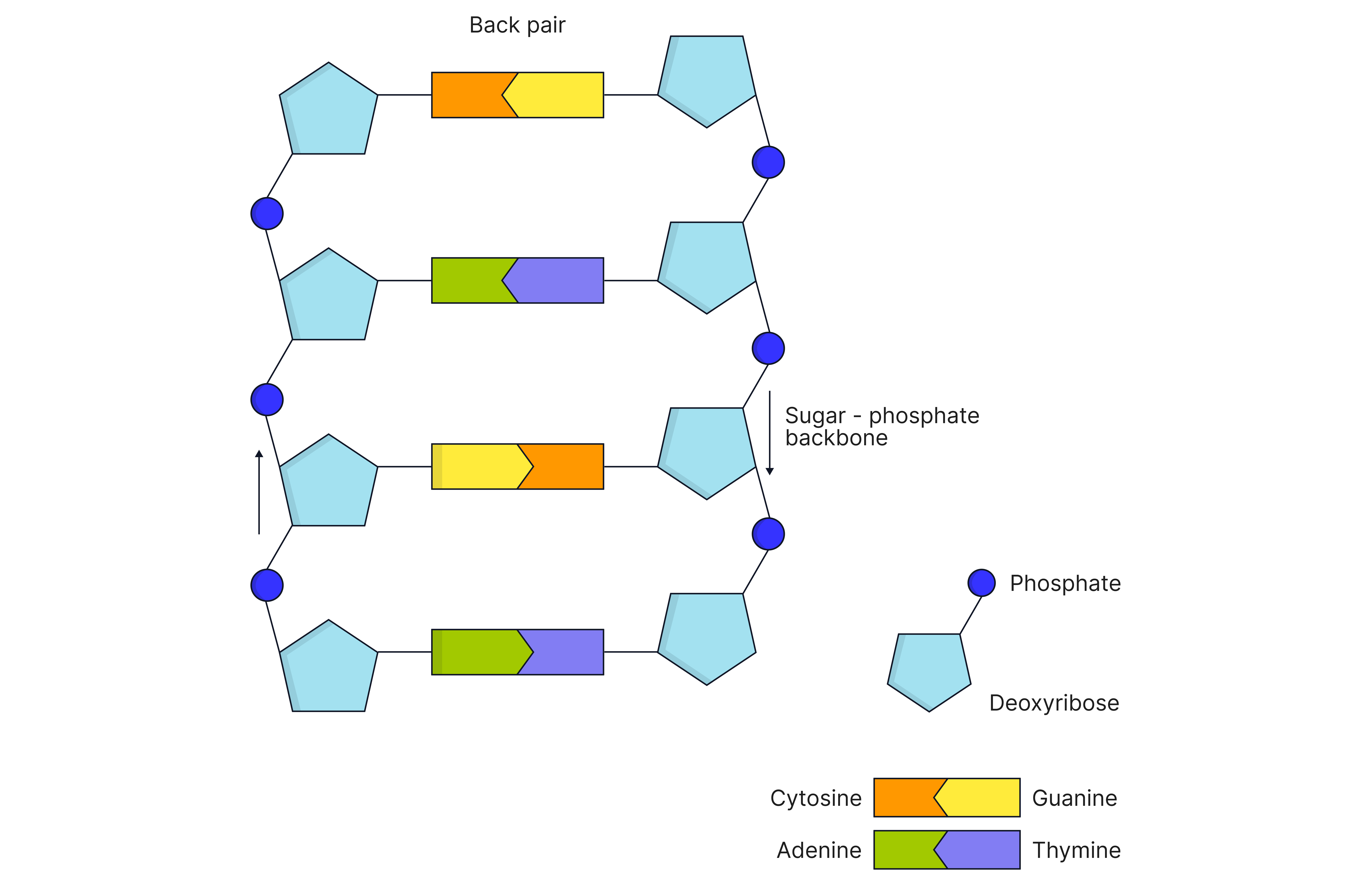
Base pairs:
Adenine and Thymine (2 hydrogen bonds)

Cytosine and Guanine (3 hydrogen bonds)

Hydrogen bonds are formed between lone pairs on O or N atoms and atoms on the base of the neighbouring strand.
Cisplatin and DNA
Cisplatin is a platinum-based anti-cancer drug that binds to DNA, disrupting replication, therefore slowing the spread of cancer.
Mode of Action:
Cisplatin forms coordinate bonds with the nitrogen atoms on guanine bases
This distorts DNA shape, preventing replication and transcription
Ethical Issues Around Cisplatin
Benefits: Treats cancer effectively
Risks: Causes side effects like nausea, hair loss, kidney damage
Ethical balance: Patient consent, quality of life, long-term risks
Practice Questions
A section of a single strand of DNA has the following sequence of bases:
A T T G C A
a) Give the sequence of bases on the complementary strand.
b) State the total number of hydrogen bonds in this section of DNA.
Answer
a) T A A C G T
b) 14 (2 + 2 + 2 + 3 + 3 + 2)
Explain how two DNA strands are held together.
Answer
1. Hydrogen bonds between bases.
2. A pairs with T, G pairs with C.
3. Specific complementary base pairing.
Describe the role of cisplatin in cancer treatment and how it works at the molecular level.
Answer
1. Cisplatin binds to DNA.
2. Forms coordinate bonds with nitrogen in guanine bases.
3. Prevents replication/transcription of DNA.
4. Stops cell division leading to cancer cell death.