Structure of the Ear
Brook Edgar & Hannah Shuter
Teachers


Brook Edgar
Physics
Hannah Shuter
Physics
Explainer Video
Structure of the Ear
The ear is comprised of three parts, the outer ear, the middle ear and the inner ear.
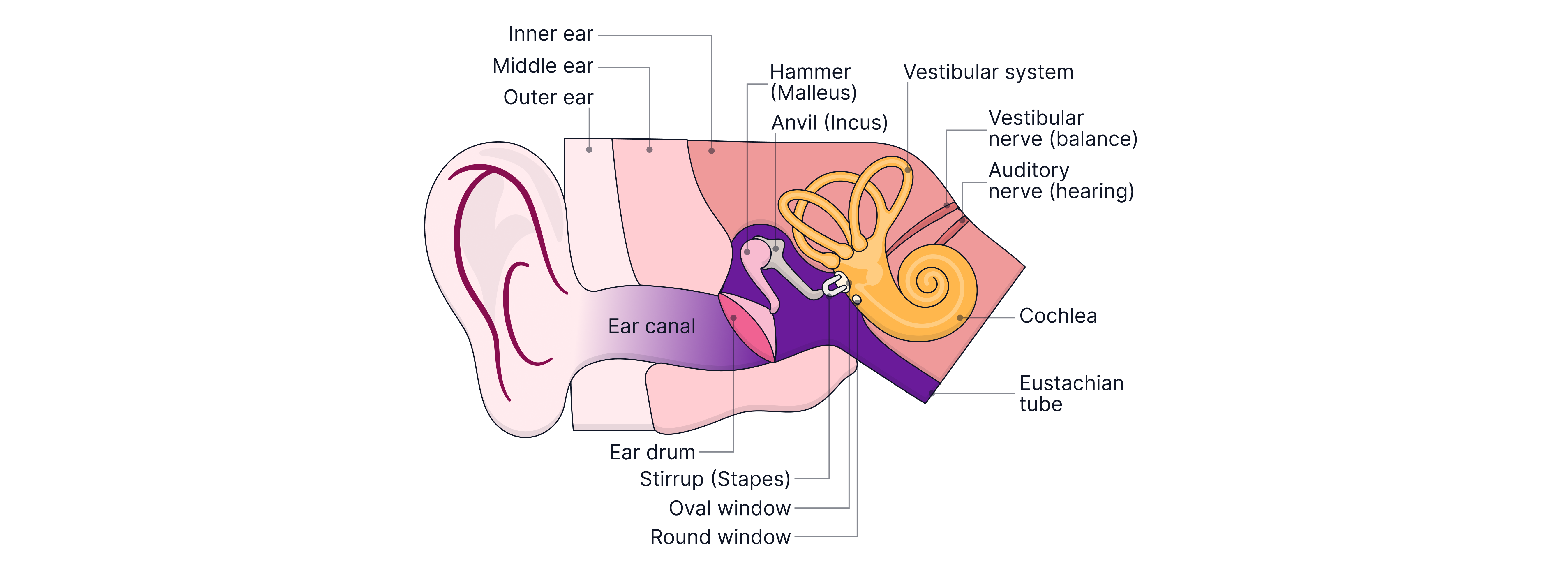
Outer ear
The ear canal funnels sound waves towards the tympanic membrane (eardrum) which vibrates when sound waves arrive. The eardrum has a diameter of approximately , and converts pressure variations in air into mechanical vibrations.
The ear canal acts as a resonant tube, naturally amplifying sounds with frequencies in the range . This resonance contributes to the ear’s maximum sensitivity around .
Middle ear
The middle ear transmits sound energy from the eardrum (tympanic membrane) to the oval window, which is the entrance to the inner ear. Vibrations are passed through three small bones called the ossicles:
Hammer (malleus)
Anvil (incus)
Stirrup (stapes)
The ossicles act as a system of levers, increasing the force at the oval window by . In addition, the eardrum has a much larger area than the oval window so the force is concentrated onto a much smaller area. Since, , the pressure at the oval window is much greater than at the eardrum (about greater).
This helps efficiently transfer sound from air into the fluid-filled inner ear.
Inner ear
The inner ear is filled with fluid and contains the cochlea, which is responsible for hearing, and the vestibular system, which is responsible for balance.
The inner ear is connected to the middle ear by two flexible membranes:
Oval window – receives vibrations from the stapes (stirrup), causing pressure waves to travel through the fluid inside the cochlea.
Round window – moves in the opposite direction to the oval window, allowing the fluid within the cochlea to move freely and preventing pressure build-up.
Worked Example:
A sound wave in the ear produces a pressure amplitude at the oval window that is times greater than the pressure amplitude at the eardrum.
Calculate the ratio of .
The area of the oval window is .
The area of the eardrum is .
Answer:
We know that the pressure on the oval window is times greater than that on the eardrum, and that :
This answer fits with what we know to be true - that the ossicles multiply the force of the sound wave by about times as it moves across the middle ear.
Worked Example:
A sound wave produces a maximum pressure change of at the eardrum.
This results in a maximum pressure change of in the fluid of the inner ear.
Explain how the ossicles help to increase the pressure transmitted to the fluid of the inner ear.
Answer:
The ossicles act as a lever system that increases the force transmitted from the eardrum to the oval window.
The force will be increased by times.
Cochlea Structure
The cochlea is a helical (spiral) cavity inside a rigid bone. It contains three fluid-filled channels:
Vestibular channel
Cochlear channel
Tympanic channel
The oval window is connected to the vestibular channel, which at its apex joins the tympanic channel, which is attached to the round window. The image below shows an "uncoiled" cochlea.

Both vestibular and tympanic channels are filled with fluid, which transmits pressure waves from the vibrations of the oval window.
The cochlear channel contains a fluid called endolymph.
The Corti
The cochlear channel contains the organ of Corti, which consists of hair cells and supporting cells resting on the basilar membrane. The basilar membrane separates the cochlear channel from the tympanic channel below. The hair cells have cilia projecting into the endolymph. When the basilar membrane vibrates, the cilia bend, generating nerve impulses in the auditory nerve that travel to the brain and are interpreted as sound.
The Sequence of Hearing
Frequency Detection in the Cochlea
Different frequencies vibrate different regions of the basilar membrane.
High frequency sounds are detected at the base of the cochlea, near the oval window, where the basilar membrane is thick and stiff.
Low frequency sounds are detected at the apex of the cochlea, where the basilar membrane is thin and flexible.
Worked Example:
Explain how the position of maximum vibration along the basilar membrane depends on the frequency of the sound.
Answer:
The basilar membrane varies in stiffness along its length. It is stiffer near the oval window and less stiff towards the apex.
High-frequency sounds produce maximum vibration near the base of the cochlea, close to the oval window.
Low-frequency sounds produce maximum vibration further along the basilar membrane towards the apex.
Worked Example:
Describe how vibrations entering the cochlea lead to the stimulation of hair cells.
Answer:
Vibrations of the oval window produce pressure waves in the fluid inside the vestibular channel. These pressure waves cause the basilar membrane to vibrate. Movement of the basilar membrane vibrate the cilia, stimulating nervous impulses.
Practice Questions
A sound wave causes a peak pressure change of at the tympanic membrane. This results in a peak pressure change of in the fluid of the inner ear.
The tympanic membrane of the ear may be treated as a circular surface with a diameter of .
Calculate the area of the oval window.
-> Check out Hannah's video explanation for more help.
Answer:
Sound waves entering the ear must be efficiently transferred from air to the fluid of the cochlea.
Describe how the structure of the ear allows sound waves to be transmitted from the ear canal to the cochlea.
You should refer to at least two different parts of the ear in your answer.
-> Check out Hannah's video explanation for more help.
Answer:
Sound waves travel along the ear canal to the tympanic membrane (eardrum), causing it to vibrate.
Vibrations are transferred to the ossicles (malleus, incus, stapes), which transmit the vibrations across the middle ear.
The stapes vibrates the oval window, producing pressure waves in the fluid of the cochlea.
Pressure waves in the cochlear fluid cause the basilar membrane to vibrate, leading to stimulation of hair cells (which then generate nerve impulses).