Sliding Filament Theory
Laura Armstrong
Teacher

Contents
Recall Questions
This topic requires prior knowledge of the structure of skeletal muscle. You can test your knowledge on this below.
What ion is stored in the sarcoplasmic reticulum and released during muscle contraction?
Calcium ions (Ca²⁺).
What is the role of ATP in cells?
Provides energy for cellular processes via hydrolysis.
Which proteins interact during muscle contraction?
Actin and myosin.
Topic Explainer Video
What Is the Sliding Filament Model?
The sliding filament model explains how muscles contract by describing how actin and myosin filaments slide past each other within a sarcomere, shortening its length and producing muscle contraction.
Important:
- Filaments do NOT shorten, they slide past one another.
- This shortens the sarcomere → shortens the myofibril → shortens the muscle.
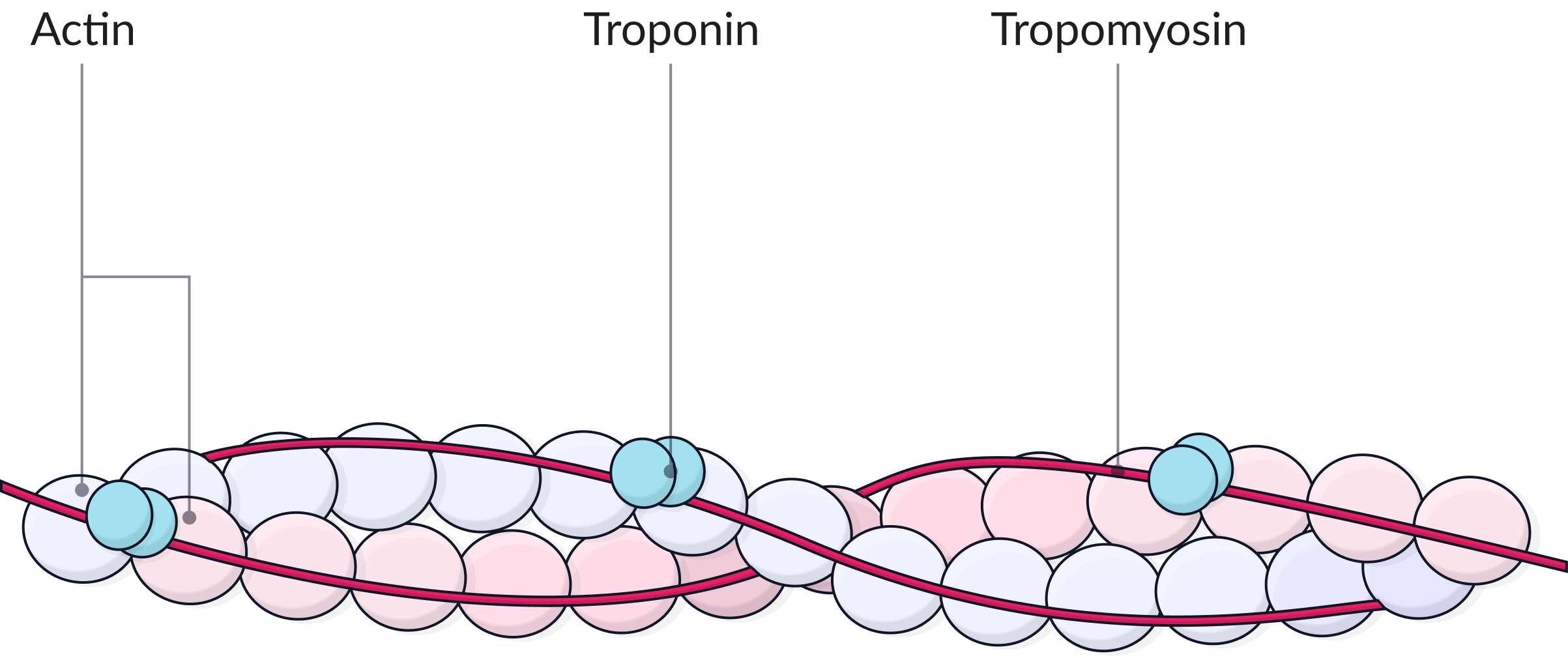
Key Structural Proteins Involved
| Protein | Function |
| Actin | Thin filament with binding sites for myosin heads. |
| Myosin | Thick filament with protruding heads that bind and pull actin inwards. |
| Tropomyosin | Blocks binding sites on actin when muscle is relaxed. |
| Troponin | Binds calcium and moves tropomyosin to expose binding sites. |
Steps in the Sliding Filament Mechanism
1. Stimulation
- Action potential reaches neuromuscular junction.
- Sarcolemma depolarises.
- T-tubules transmit impulse deep into muscle fibre.
- Ca²⁺ ions released from sarcoplasmic reticulum into sarcoplasm.
2. Binding Site Exposure
- Ca²⁺ binds to troponin.
- Troponin changes shape: pulls tropomyosin away from actin’s binding sites.
- Myosin heads can now attach to actin: forms an actin-myosin cross-bridge.
3. Power Stroke
- ADP + Pi are released from myosin head.
- Myosin head tilts, pulling actin filament inwards towards the centre of sarcomere.
- This is the power stroke that causes sarcomere shortening.
4. Detachment
-
ATP binds to myosin head → causes it to detach from actin (the actin-myosin crossbridges break).
5. Re-cocking
- ATP is hydrolysed to ADP + Pi by ATPase.
- Myosin head returns to its original “cocked” position ready for another cycle.
- They will then reattach to the actin filament further along.
This cycle repeats many times during a single contraction as long as Ca²⁺ and ATP are present. There are also multiple myosin heads pulling on the actin filaments, this increases the force of contraction.
After contraction, calcium ions are actively transported back into the sarcoplasmic reticulum. This requires energy from ATP.

Changes in the Sarcomere During Contraction
| Structure | What Happens During Contraction |
| A-band | Stays the same (length of myosin). |
| I-band | Shortens. |
| H-zone | Shortens. |
| Z-lines | Move closer together. |
| Sarcomere | Shortens overall. |
Key Terms
- Cross-bridge: Connection formed when myosin head binds to actin.
- Power stroke: Movement of myosin head pulling actin inwards.
- ATPase: Enzyme that hydrolyses ATP for energy.
- Sarcomere: Basic unit of contraction; Z-line to Z-line.
- Tropomyosin & Troponin: Regulatory proteins controlling binding site access.
No answer provided.
Exam Tips
Avoid saying “myosin moves” — instead say “myosin pulls actin” or “actin filaments slide past myosin”.
Always state ATP is needed to break the actin-myosin crossbridge and to reset the myosin head.
Be clear on the role of Ca2⁺, to expose the myosin binding sites on actin.
No answer provided.
Describe how the sliding filament model explains muscle contraction. (6 marks)
- Action potential causes Ca²⁺ to be released from the sarcoplasmic reticulum.
- Ca²⁺ binds to troponin, causing tropomyosin to move and expose actin binding sites.
- Myosin heads bind to actin forming cross-bridges.
- ADP and Pi are released from the myosin head, causing the head to tilt, pulling actin inwards.
- ATP binds to myosin head, causing it to detach from actin.
- ATP is hydrolysed to ADP + Pi, re-cocking the myosin head
- This cycle repeats, causing filaments to slide and sarcomeres to shorten.
Practice Question 1
Try to answer the practice question from the TikTok on your own, then watch the video to see how well you did!
Practice Question 2
If you want to try out another one, check this video out and see how you do!