Light microscopes and electron microscopes
Laura Armstrong & Joe Wolfensohn
Teachers


Recall Questions
This topic requires prior knowledge of the structure and function of cells and organelles and using a light microscope.
What does a microscope do?
They magnify small objects (such as cells) so they can be seen in more detail.
What is magnification?
How many times larger the image is than the actual object.
What is resolution?
The ability to distinguish between two separate objects.
Topic Explainer Video
Check out this @JoeDoesBiology video that explains light microscopes and electron microscopes, then read the study notes. Once you’ve gone through them, don’t forget to try the practice questions!
Light vs Electron Microscopes
Microscopes are used to view cells and cell structures. There are two main types, light and electron.
Light Microscopes (Optical Microscopes)
Key Features:
-
Use light and lenses to magnify.
-
Can view living or dead specimens.
-
Magnification: Up to ×1500.
-
Resolution: ~200 nm.
-
Produce colour images.
Used for:
Viewing whole cells, tissues, and living processes.
Electron Microscopes
-
Electrons pass through the specimen.
-
Vacuum required so the specimen must be dead.
-
Higher resolution: ~0.1 nm.
-
Higher magnification.
-
Detailed images of internal structures.
-
Produce black and white images only.
-
Using an electron microscope requires specialist skills and the preparation process is complex and time consuming.
Used for:
Studying sub-cellular structures (e.g. organelles) or very small specimens such as viruses.
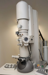
An electron microscope.
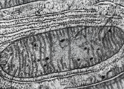
An image of a mitochondrion using an electron microscope.
Comparison Table
|
Feature |
Light Microscope |
Electron Microscope |
|
Radiation Used |
Light |
Electrons |
|
Resolution |
Lower |
Higher |
|
Magnification |
Lower) |
Higher |
|
Can View Live Cells? |
Yes |
No (vacuum required) |
|
Colour Image |
Yes (with stain) |
No (black & white only) |
|
Cost and Size |
Low cost, portable |
Expensive, large |
Key Terms
-
Resolution – the ability to distinguish between two separate objects.
-
Magnification – how much larger the image appears compared to real size.
No answer provided.
Exam Tip
If a question asks for comparison, write in clear contrasts (e.g. Light microscopes give colour images whereas electron microscopes only produce black and white images).
No answer provided.
Practice Question
Describe three differences between using a light microscope and an electron microscope. (3 marks)
Any 3 from
-
A light microscope uses light, whereas an electron microscope uses electrons.
-
A light microscope has a lower resolution than an electron microscope.
-
A light microscope has a lower magnification than an electron microscope.
-
A light microscope can be used to study living specimens whereas an electron microscope can only be used to study dead specimens.
-
Light microscopes give colour images whereas electron microscopes only produce black and white images.
More Practice
Try to answer these practice questions from the TikTok videos on your own, then watch the videos to see how well you did!