Tissue fluid
Laura Armstrong & Joe Wolfensohn
Teachers


Recall Questions
This topic requires prior knowledge of osmosis, pressure gradients and the structure of blood vessels. You can test your knowledge on these below.
What is plasma, and what does it contain?
Plasma is the liquid component of blood, containing water, proteins, ions, nutrients, hormones, and waste products.
What is the function of capillaries in exchange of substances?
Capillaries allow the exchange of oxygen, nutrients, and waste products between the blood and surrounding tissues due to their thin walls and small size.
What is osmosis?
The diffusion of water from a higher water potential to a lower water potential, across a partially permeable membrane
Topic Explainer Video
Check out this @JoeDoesBiology video that explains tissue fluid or read the full notes below. Once you've gone through the whole note, try out the practice questions!
Intro to Tissue Fluid
Tissue fluid bathes almost all cells in our body that are outside of our circulatory system. Tissue fluid is formed at the arteriole end of the capillaries.
Blood flows in our arteries before moving into narrower arterioles and then into capillary networks. It is in the capillaries that exchange of substances can take place (as the walls of capillaries consist of a single layer of endothelial cells).
Substances are not exchanged directly from capillaries to cells. The substances pass out of the capillaries and form what we call tissue fluid. It is from this tissue fluid that cells can then obtain the substances they need, such as glucose and oxygen for respiration.
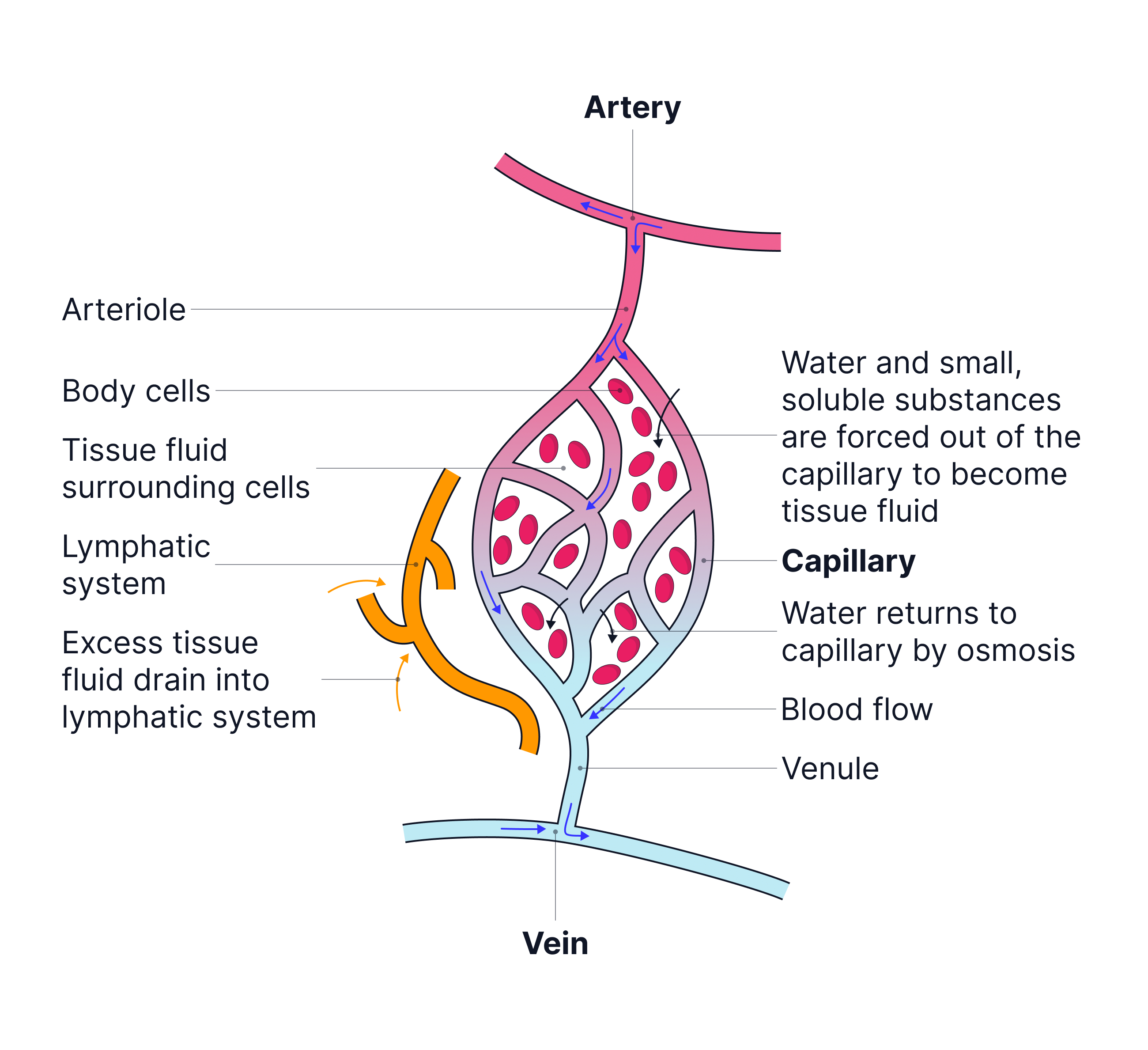
At the venule end of the capillary network, water moves back into the capillaries from the tissue fluid by osmosis. The rest of the tissue fluid, including waste products made by our cells, drains into lymphatic vessels before it returns to the circulatory system.
The formation of tissue fluid
1. At the arteriole end there is higher hydrostatic pressure (blood pressure). This is generated by the strong contractions of the heart muscle in the left ventricle.
This hydrostatic pressure is high enough that it forces fluid out of the capillaries.
This fluid will contain water, oxygen, glucose, amino acids, fatty acids and mineral ions as these substances are small enough to pass through the wall (endothelium) of the capillary.

2. These substances form tissue fluid and will surround the cells. From this tissue fluid, oxygen and glucose can then diffuse into cells to be used in respiration, as well as any amino acids, fatty acids or mineral ions the cells may need.
Large proteins, as well as red blood cells, will remain in the capillaries as they are too large to pass through the walls of the capillaries.
The proteins that remain in the blood plasma will lower the water potential of the blood. This creates a water potential gradient between the blood in the capillary and the tissue fluid. In turn, this creates an osmotic pressure, acting against the hydrostatic pressure.
However, as the hydrostatic pressure is greater than the opposing osmotic pressure at the arteriole end, the net movement of water is out of the capillaries and into the tissue fluid.
The return of tissue fluid
1. At the venule end the hydrostatic pressure is reduced. This is because:
- The blood has slowed down as it has travelled through the narrow capillaries and experienced friction from the capillary walls.
- The blood is now further away from the heart.
- The blood has lost volume as it has moved along the capillary due to the loss of water into the tissue fluid.
2. The large proteins that remain in the blood are lowering the water potential in the capillary and are creating a water potential gradient between the blood in the capillary and the tissue fluid.
3. At the venule end, the osmotic pressure created by this water potential gradient is greater than the opposing hydrostatic pressure (which has been lowered).
Therefore, water will move back into the capillaries from the tissue fluid by osmosis, down the water potential gradient.
4. Excess tissue fluid will drain into the lymphatic system where we call the fluid ‘lymph’. It enters lymph vessels and is carried back into the circulatory system via a vein in the chest called the subclavian vein.
Lymph moves due to the contraction of body muscles that squeeze the lymph vessels in the direction of the heart. Any backflow of lymph is prevented by valves.
If a person has high blood pressure, also known as hypertension, the hydrostatic pressure is even greater at the arteriole end of the capillary networks. This pushes more fluid out of the capillaries and leads to the formation of more tissue fluid. Tissue fluid may begin to accumulate around their body cells and cause swelling known as oedema.
Key Terms
-
Hydrostatic Pressure: The pressure exerted by blood against capillary walls, forcing fluid out.
-
Oncotic Pressure: The osmotic pressure due to plasma proteins reducing the water potential, pulling water back into capillaries.
-
Lymph: The fluid inside lymphatic vessels, derived from tissue fluid.
- Lymphatic System: A network of vessels that drain excess tissue fluid and help in immune responses.
No answer provided.
Exam Tip
When describing the movement of water back into the capillaries at the venule end, always say it moves by osmosis.
No answer provided.
Describe how tissue fluid is formed and how it is returned to your circulatory system. (6 marks)
Formation
1. High blood pressure or hydrostatic pressure
2. Forces water / fluid out of the capillaries (at the arteriole end)
3. Large proteins will remain in the capillary
Return
4. There is a lower water potential in the capillary (at the venule end)
5. Due to (plasma/ large) proteins (which have remained in the blood)
6. Water enters the capillary
7. By osmosis
8. Excess tissue fluid drains into the lymphatic system
Practice Question 1
Try to answer the practice question from the TikTok on your own, then watch the video to see how well you did!
Practice Question 2
If you want to try out another one, check this video out and see how you do!