Required practical - using a light microscope and doing a biological drawing
Laura Armstrong & Joe Wolfensohn
Teachers


Recall Questions
This topic requires prior knowledge of cell structures.
Which part of the cell releases energy during respiration?
Mitochondria
Name 3 structures found in a plant cell that are not found in an animal cell?
Cell wall, chloroplast, large vacuole
In which part of the cell do chemical reactions take place?
Cytoplasm
Topic Explainer Video
Check out these @JoeDoesBiology videos that explain how to use a light microscope or read the full notes below. Once you've gone through the whole note, try out the practice questions!
Required Practical – Microscopy
Cells cannot be seen with the naked eye. Microscopes produce an image of the cell that is larger than the actual cell. The magnification is how many times larger the image is. Some sub-cellular structures may still not be visible. The resolution of the microscope is how easy it is to distinguish between two points that are close together. Light microscopes have a low resolution so ribosomes, for example, would not be visible.
Aim of the practical:
To observe plant cells using a light microscope.
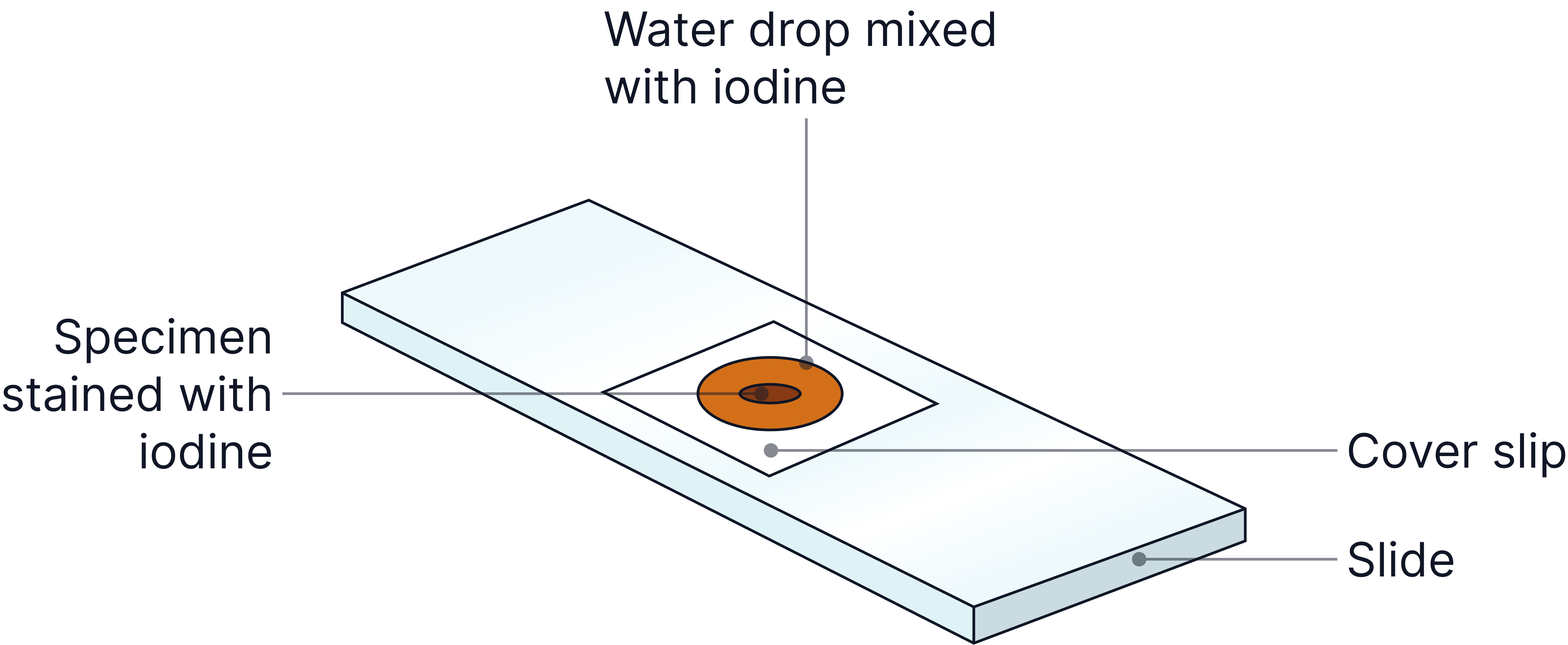
Step 1: Preparing the Slide (Onion Cells Example)
-
Peel a thin layer from an onion.
-
Place the tissue flat on a clean microscope slide.
-
Add a drop of iodine solution (stains the cell structures to make them visible).
-
Use a mounted needle to carefully lower a cover slip at an angle over the sample – this helps to avoid air bubbles!
The specimen needs to be thin so that light can pass through it. Ideally it will be a single layer of cells.
The specimen also needs staining to provide contrast so that the sub-cellular structures are visible.
No answer provided.
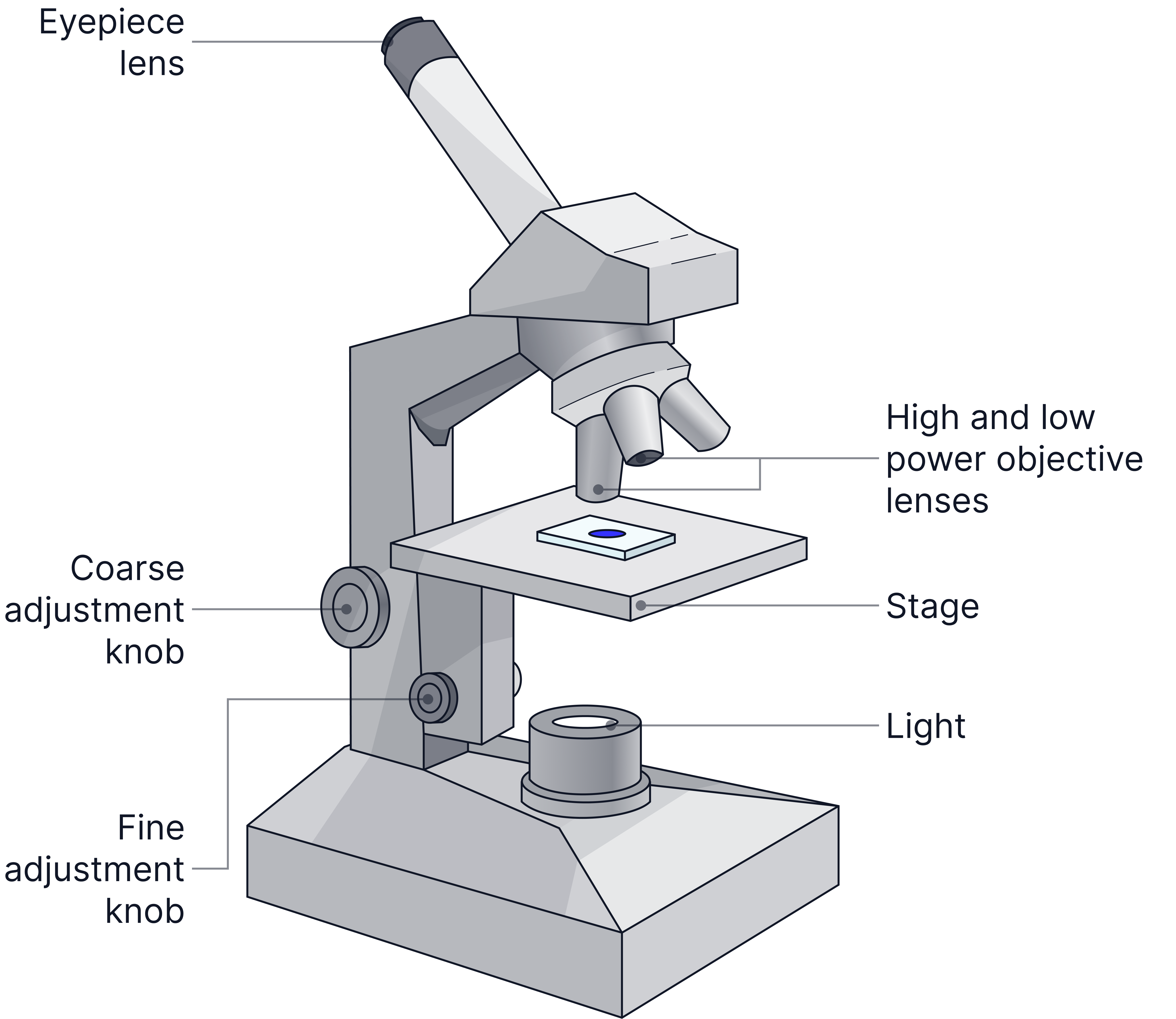
Step 2: Using the Microscope
-
Start with the lowest magnification objective lens. This provides the widest field of view to know which area to increase the magnification on.
-
Use the coarse focus knob to bring the slide into general focus.
-
Use the fine focus knob to sharpen the image.
-
Switch to a higher magnification objective lens if needed and refocus using only the fine focus knob.
-
Calculate total magnification:
Total Magnification = Eyepiece Lens magnification × Objective Lens magnification
E.g if the eyepiece lens is x5 and the objective lens is x40 the total magnification will be 5 x 40 = 200.
No answer provided.
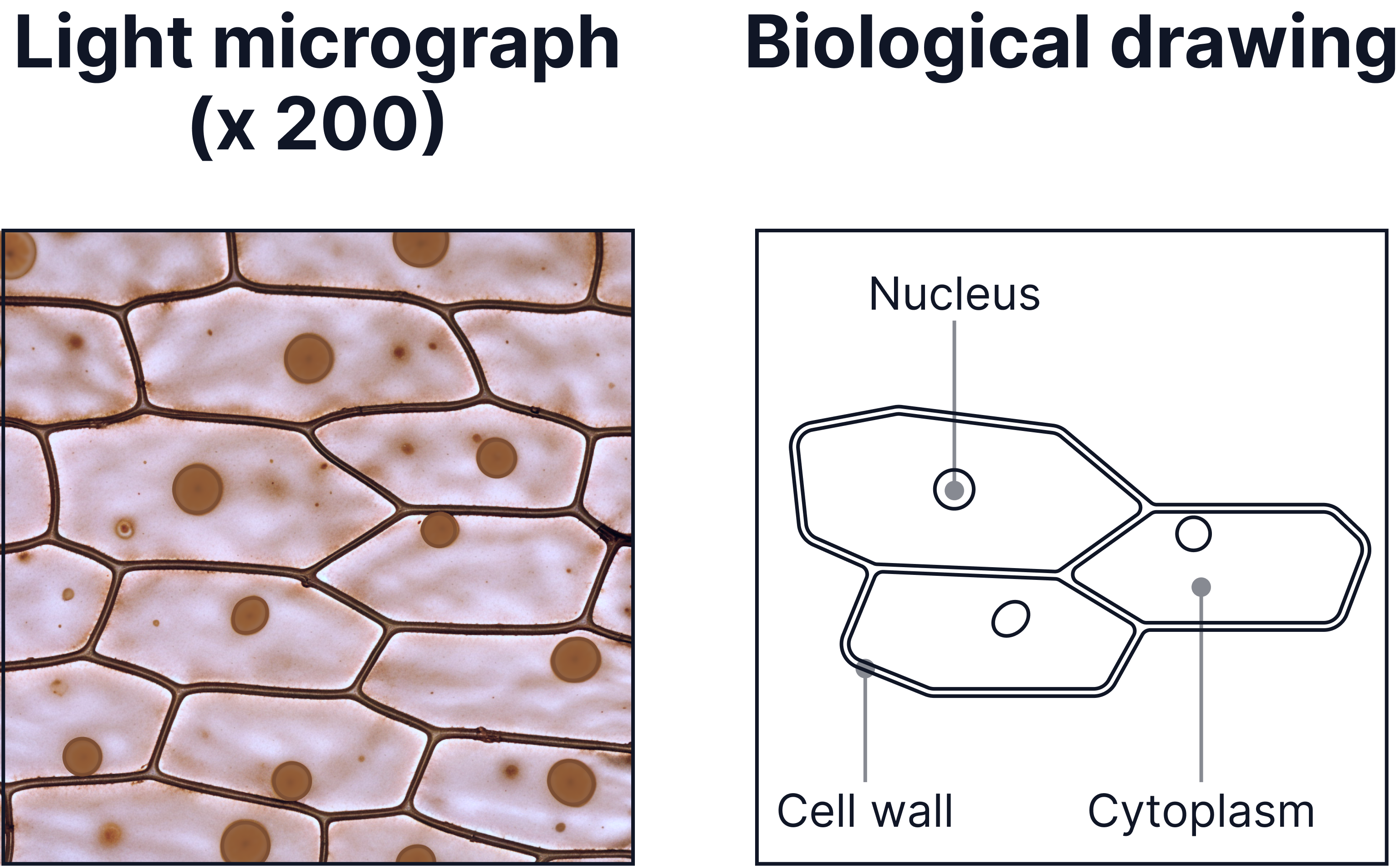
Step 3: Biological Drawing
A biological drawing is used to represent what you can see in the image.
-
Use a sharp pencil, draw only what you see clearly.
-
Use continuous single lines, lines must not be broken.
-
Label the structures you can see.
-
Use ruled label lines.
-
No shading or colouring.
-
Include a title and the magnification used.
Example
No answer provided.
Risk Assessment
|
Risk |
Hazard |
Control Measure |
|
Using a scalpel |
Sharp scalpel blades can cause cuts when slicing onion skin. |
Always cut away from your fingers. Cut on a hard surface such as on a cutting tile. |
|
Using a mounted needle |
Pointed mounted needle can cause injury if misused. |
Always point the sharp end away from hands and others. |
|
Using iodine solution |
Iodine can stain skin and irritate eyes. It is an irritant. |
Wear safety goggles so iodine does not enter the eyes. Wash off any splashes on skin immediately with water. |
|
Slips from spills |
Dropped slides, water, or iodine can make the bench or floor slippery. |
Wipe up spills straight away. Keep the work area tidy and dry. |
Key Terms
- Magnification – how much larger the image is than the real object.
- Resolution – how easily you can distinguish between separate objects that are close together.
No answer provided.
Exam Tips
- Don't use the word zoom when defining magnification, make sure to use the definition given above.
- When using a microscope, increase the magnification if the image is too small, focus the image if it is blurry or unclear.
No answer provided.
Practice Question
A student prepared a slide of onion cells using iodine and observed it under a microscope. Explain why they used a sharp scalpel and iodine. (3 marks)
-
Used a scalpel to make sure the specimen / sample was thin / a single layer of cells,
-
So light could pass through.
-
Used iodine to stain the sub-cellular structures, so they can be seen.
More Practice
Try to answer these practice questions from the TikTok videos on your own, then watch the videos to see how well you did!