Reflex actions
Laura Armstrong & Joe Wolfensohn
Teachers


Contents
Recall Questions
This topic requires prior knowledge of homeostasis and negative feedback.
What is negative feedback?
A change in a condition triggers a response that counteracts (reverses) the change to restore normal conditions.
What is a co-ordinator in the nervous system?
The central nervous system (brain and spinal cord), which processes information and coordinates a response.
What is an effector?
A muscle or gland that carries out a response to a stimulus (e.g. contracts or secretes hormones).
Topic Explainer Video
Check out these @JoeDoesBiology and @Lauradoesbiology videos that explain reflex actions, then read the study notes. Once you’ve gone through them, don’t forget to try the practice questions!
What is a Reflex Action?
- A reflex action is an automatic and rapid response to a stimulus.
-
It helps protect the body tissues from damage (e.g. pulling your hand away from something hot to prevent burning).
-
Reflexes do not involve the conscious part of the brain.
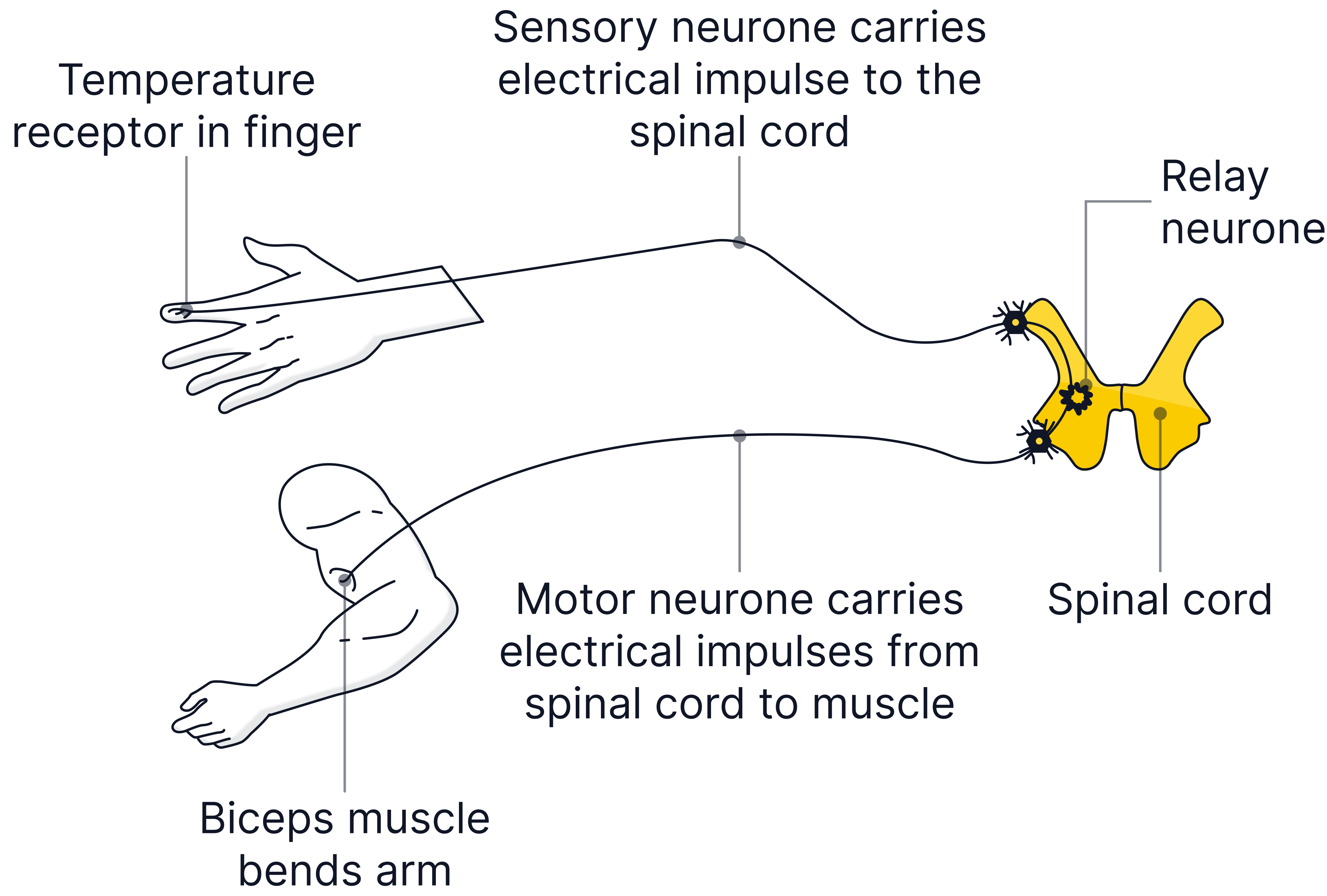
Reflex Arc Pathway
-
Stimulus – a change in the environment e.g. an increase in temperature.
-
Receptor – detects the stimulus e.g. temperature receptors in the skin.
-
Sensory neurone – carries electrical impulse to the central nervous system (CNS).
-
Relay neurone – in the CNS (usually in the spinal cord), it connects the sensory neurone to a motor neurone.
-
Motor neurone – carries electrical impulse to an effector.
-
Effector – a muscle or a gland that causes a response - muscles contract or glands secrete a chemical substance (such as a hormone).
-
Response – e.g. hand pulls away from the hot pan.
Key idea: Reflexes skip the brain’s conscious areas to allow for faster responses.
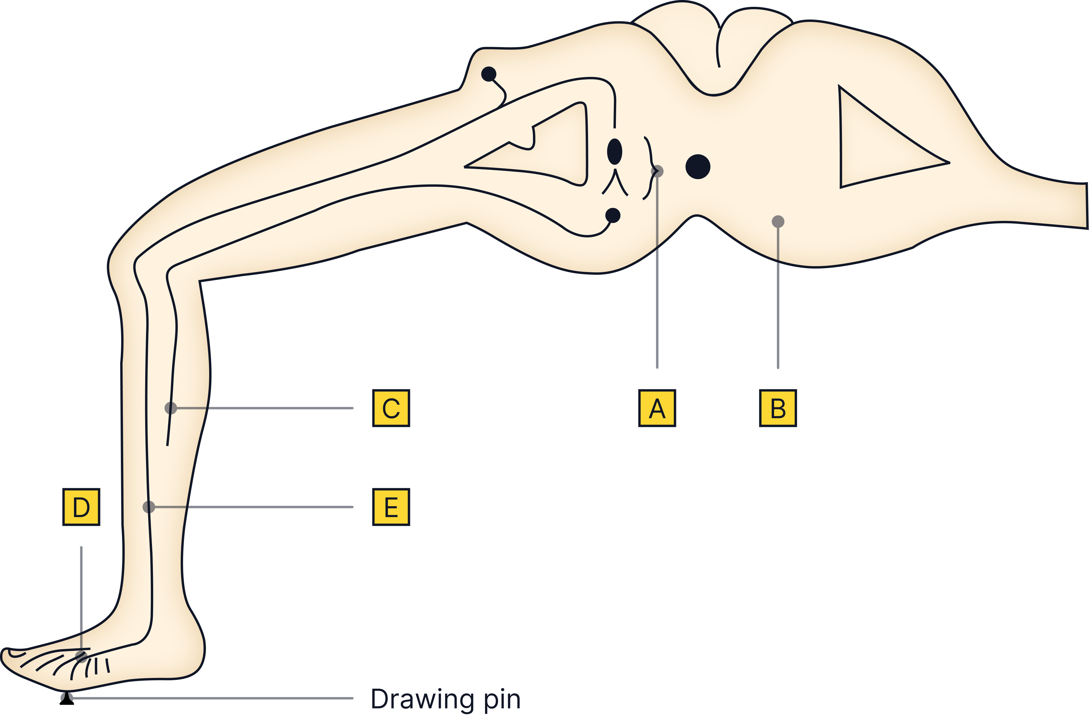
Reflex arc diagrams
Check if you can label the reflex arcs given below correctly!
-
The relay neurone
-
The spinal cord
-
The motor neurone
-
The receptors
-
The sensory neurone

-
The spinal cord
-
The relay neurone
-
The receptors
-
The sensory neurone
-
The effector (muscle)
-
The motor neurone
Synapses
-
A synapse is the gap between two neurones.
-
The electrical impulse triggers the release of chemical messengers (neurotransmitters).
-
These diffuse across the synapse and bind to the next neurone, starting a new electrical impulse.
-
The diffusion of a chemical is slower than the transmission of an electrical impulse so synapses slow the reflex action down slightly.
-
Synapses are found between the sensory neurone and the relay neurone, the relay neurone and the motor neurone, and between the motor neurone and the effector.
Key Terms
- Reflex - An automatic, rapid response to a stimulus.
- Receptor - Cells that detect a stimulus and initiate an electrical impulse along a sensory neurone.
- Sensory neurone - Carries electrical impulses from receptors to the CNS.
- Relay neurone - Found in the CNS; connects sensory and motor neurones.
- Motor neurone - Carries electrical impulses from the CNS to effectors.
- Synapse - A tiny gap between neurones where chemicals diffuse to transmit the signal.
- Effector - Muscle or gland that carries out the response.
No answer provided.
Exam Tips
Use the correct order in reflex questions – receptor → sensory neurone → relay neurone → motor neurone → effector → response.
Always say neurones transmit ‘electrical impulses’ not messages or signals!
No answer provided.
Practice Questions
Question 1
A student accidentally touches a sharp object.
Her hand is immediately pulled away from the object.
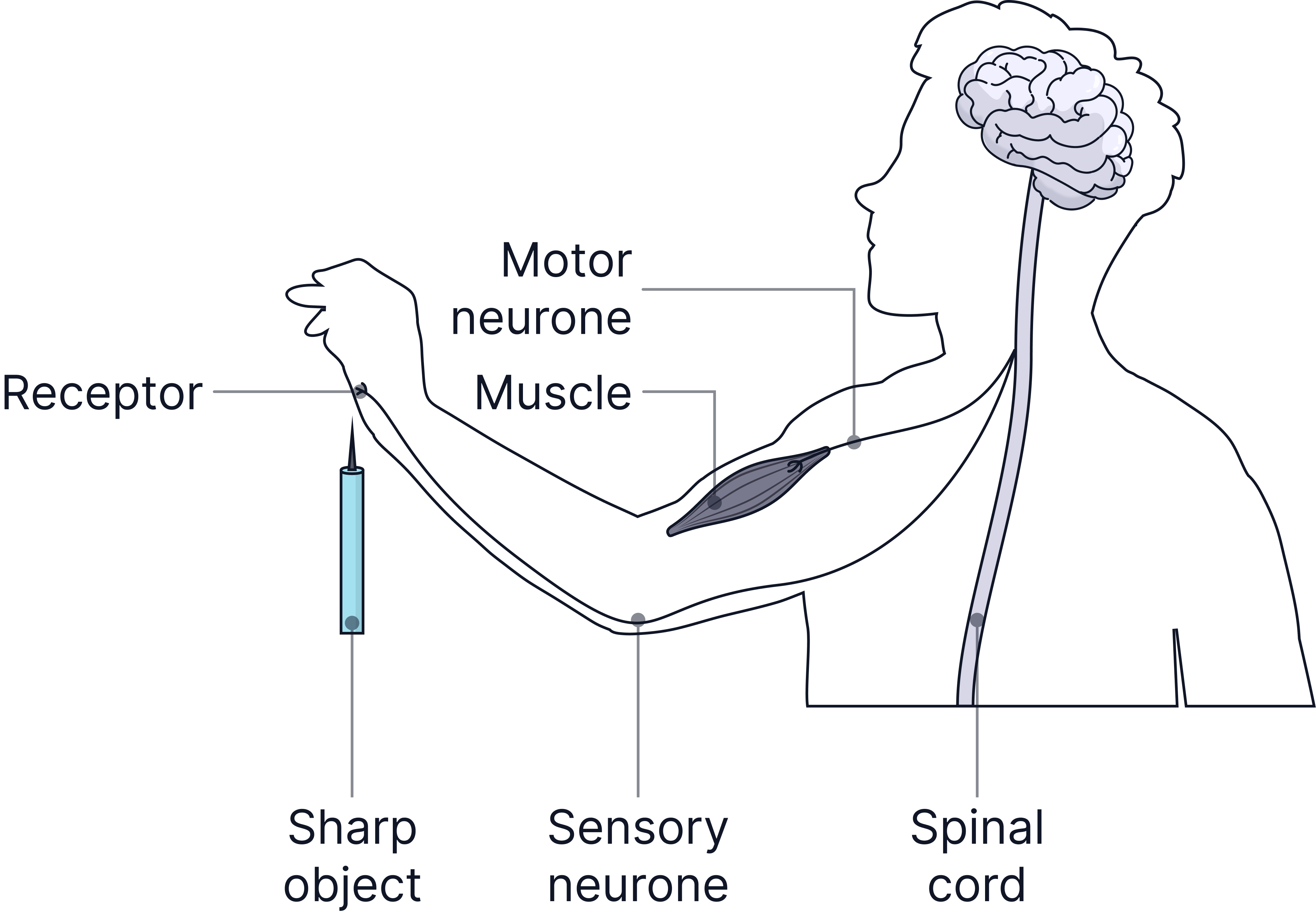
Describe how the structures labelled on the diagram are involved in this reflex action. (4 marks)
-
receptor detects stimulus / sharp object.
-
electrical impulse passes along sensory neurone to spinal cord.
-
electrical impulse passes from spinal cord along motor neurone to muscle.
-
muscle contracts to move the hand / arm.
Question 2
Describe the difference between the function of a receptor and the function of an effector.
In your answer you should give one example of a receptor and one example of an effector. (4 marks)
receptors detect stimuli or convert stimulus into an electrical impulse.
example of a receptor (allow any appropriate organ or part of an organ, eg eye / retina / skin or named type of receptor eg light receptor, temperature receptor).
effectors allow / make response or convert an impulse to an action.
effector is a muscle / gland (allow an example or a named muscle or gland such as bicep or pancreas).
More Practice
Try to answer these practice questions from the TikTok videos on your own, then watch the videos to see how well you did!