Triple Science Only - The Brain
Laura Armstrong & Joe Wolfensohn
Teachers


Contents
Recall Questions
This topic requires prior knowledge of the nervous system.
What does the central nervous system consist of?
The brain and spinal cord.
How is information transmitted along neurones?
As electrical impulses.
In a reflex arc, how do we describe the role of the CNS?
It acts as the co-ordinator.
Topic Explainer Video
Check out this @Lauradoesbiology video that explains the brain, then read the study notes. Once you’ve gone through them, don’t forget to try the practice questions!
What is the brain?
What is the brain?
-
The brain is part of the central nervous system (CNS).
-
It controls complex behaviours.
-
It is made of billions of interconnected neurones.
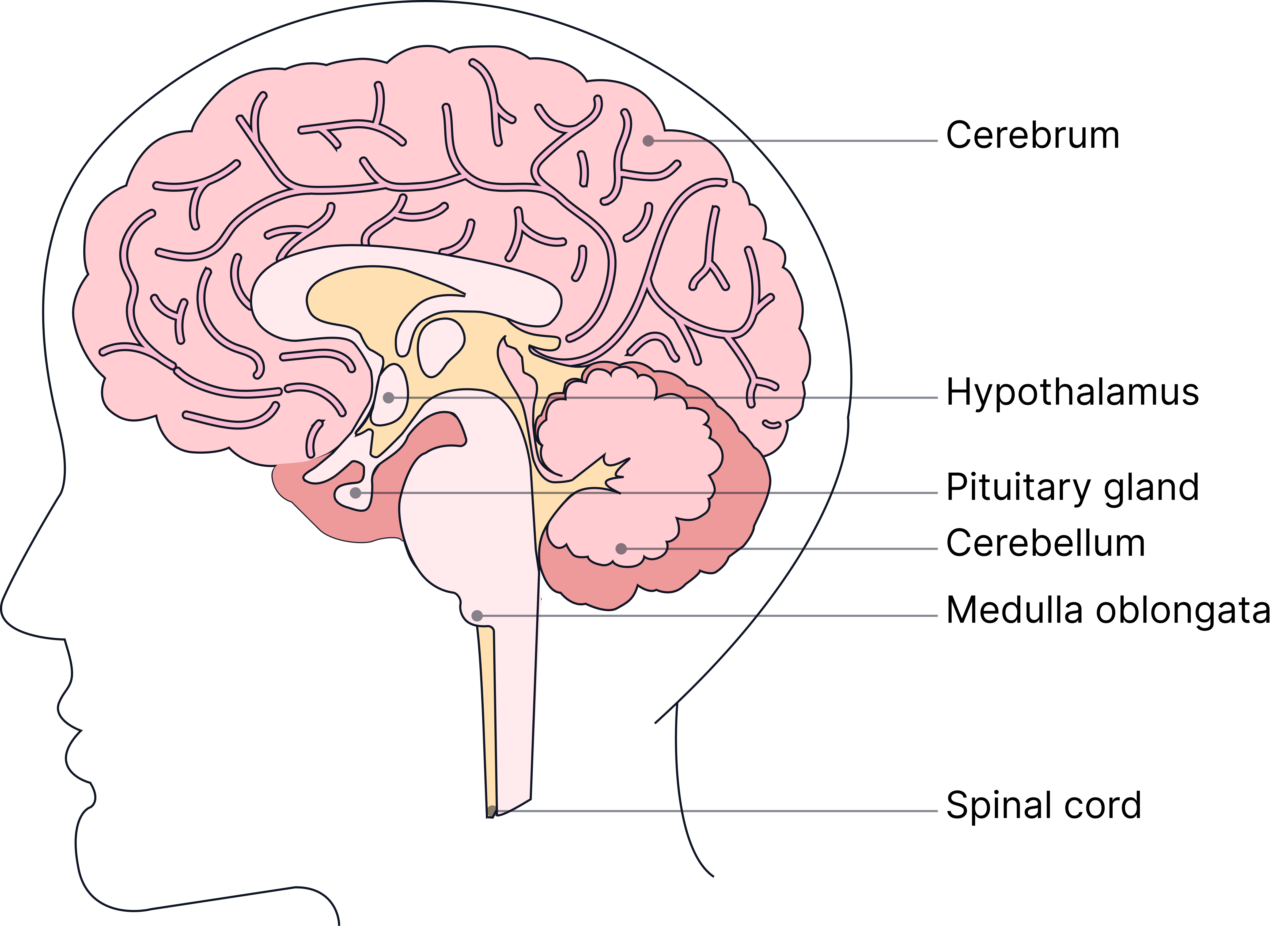
Major Parts of the Brain and Their Functions:
|
Part of the Brain |
Function |
|---|---|
|
Cerebral cortex / Cerebrum |
Controls consciousness, memory, intelligence, and language. |
|
Cerebellum |
Coordinates muscular activity and balance. |
|
Medulla |
Controls unconscious activities like heartbeat and breathing. |
|
Hypothalamus |
Regulates body temperature and helps maintain homeostasis. |
|
Pituitary gland |
The 'master gland' – releases hormones that control other glands in the endocrine system, such as FSH, LH, ADH and TSH |
Higher Tier Only
Studying the Brain
Neuroscientists are scientists who study the brain.
They can use several methods to map brain function:
-
Brain damage: Observing behavioural changes after injury to the brain.
They can study people with brain damage to learn more about what different parts of the brain do.
A well-documented example of brain damage is of Phineas Gage, who in 1848 had a serious accident whilst laying railway tracks and an iron rod went through his skull.
Phineas survived the accident, but it was documented that his personality changed following it.
It was noted that he lost his inhibitions socially and emotionally.
Doctors realised the changes in Phineas were due to the damage in the particular parts of the brain that the iron rod had passed through.
This important case allowed scientists to examine the effect of the injuries on his brain activity.
-
Electrical stimulation: Stimulating parts of the brain and monitoring the effects.
The brain can be stimulated electrically by placing electrodes on the scalp or directly on the brain.
Scientists can stimulate different parts of the brain to see what the response is.
For example, if a region of the brain controlling movement is stimulated, a patient’s muscles will contract.
They can also use electrodes to monitor electrical activity in the brain, this can help to diagnose conditions like epilepsy.
-
MRI scans: Detailed imaging of brain structure and activity.
Neuroscientists can use MRI scans to monitor the electrical activity in the brain.
Scientists can use it to see which parts of the patient’s brain are active when they do different things, such as listen to music, recall a memory or look at images which provoke emotion.
MRI scans can also be used to look for abnormalities in the brain, such as swelling, tumours and any internal bleeding or blockages in blood flow
Patients must remain as still as possible during most MRI scans, as any movement can cause the images to become blurred and unreadable.
The brain is incredibly complex, and our limited knowledge means that treating brain damage and disease is very difficult!
Brain surgery has many risks- potentially damaging the brain tissue further and leading to lifelong impact on a person’s intelligence, memory, personality or movement.
No answer provided.
Key Terms
- Cerebral cortex / Cerebrum - Outer layer of the brain that controls consciousness, memory, intelligence, and language.
- Medulla - Brain area that controls involuntary functions like breathing and heart rate.
- Cerebellum - Brain region that coordinates movement and balance.
- Hypothalamus - Part of the brain that regulates body temperature and helps maintain internal balance (homeostasis).
- Pituitary gland - The master endocrine gland – releases hormones that regulate many other glands in the body.
- MRI scan - A method used to produce detailed images of the brain to study structure and activity.
No answer provided.
Exam Tip
Use labelled diagrams to revise so you are able to locate and label the key structures of the brain.
No answer provided.
Practice Question
Describe how scientists have been able to map the functions of different areas of the brain. (3 marks)
-
By studying patients with brain damage and observing the effects on behaviour.
-
By electrically stimulating areas of the brain to observe responses.
-
By using MRI scans to see which parts of the brain are active during specific tasks.
More Practice
Try to answer the practice question from the TikTok video on your own, then watch the video to see how well you did!